Chapter 105 Abnormalities that mimic obstruction include chronic intestinal pseudoobstruction (CIPO), defined by a consensus group as a “rare, severe disabling disorder characterized by repetitive episodes or continuous symptoms and signs of bowel obstruction, including radiographic documentation of dilated bowel with air-fluid levels, in the absence of a fixed, lumen-occluding lesion.”1,2 Dilation of various parts of the gastrointestinal (GI) tract, including the small bowel, is characteristic of intestinal pseudoobstruction; these entities are further discussed in Chapter 106. Adhesions are a common cause of obstruction in patients who have undergone prior abdominal surgery. Obstruction secondary to adhesions is more common in neonates, particularly after surgery for repair of abdominal wall defects such as gastroschisis. In older children, adhesive obstruction occurs most often after pelvic surgery, usually after appendectomy.3 The postappendectomy incidence does not seem to be related to positive versus negative appendectomy, but some data suggest that it is decreased after a laparoscopic versus an open procedure.4 Several types of hernias can lead to bowel obstruction in children, and overall they are responsible for approximately 10% of bowel obstructions.3 Inguinal hernias are a relatively common condition in children, with an overall incidence between 0.8% and 4.4%.5 The vast majority of inguinal hernias in children are indirect hernias related to a patent processus vaginalis.6 The incidence of inguinal hernia is 10 times higher in boys than in girls.7 Inguinal hernias have both a higher incidence (~ 30%) and a higher risk of incarceration (31%) in premature infants.8 Other hernias that can lead to SBO include mesocolic (right or left paraduodenal) hernias (see Chapter 103) and herniation of bowel around an incompletely fixated falciform ligament. Omphalomesenteric remnants are the residua of the embryologic yolk stalk, and they become apparent in several ways, depending on the type of abnormality and ensuing complications (see also Chapter 103). Normally, the entire attachment regresses. The lumen may be obliterated, but a fibrous cord may persist, with or without cystic remnants within it. Attachment of the mid small intestine to the anterior abdominal wall via a persistent cord may lead to distal SBO in a manner similar to an adhesion; or the remnant may predispose to volvulus of a bowel loop about the fibrous cord. Adynamic ileus can cause bowel distension and may mimic mechanical obstruction. Adynamic ileus occurs invariably following major abdominal operations, and if prolonged, it may be difficult to differentiate from an early postoperative or recurrent obstruction.3 In addition, adynamic ileus may also occur in the setting of burns, trauma, and certain medications. The pathophysiology of postoperative ileus remains incompletely understood, but the disruption of the normal autonomic gastrointestinal (GI) motility is multifactorial and related to pharmacologic, inflammatory, hormonal, metabolic, and neuropsychologic influences.9 The degree of abdominal distension depends on the level of obstruction; more distal levels are associated with greater distension. SBO in children usually presents with persistent bilious vomiting and abdominal pain.10 Infants and young children may also come to medical attention with irritability and refusal to eat. The presence of fever is concerning for ischemia and bowel compromise, particularly if accompanied by unremitting pain unresponsive to proximal decompression. Patients with long-standing complete obstruction will demonstrate dilated loops of bowel with differential air-fluid levels on horizontal-beam images and absence of stool and gas in the colon (Fig. 105-1, A and B). In partial obstruction, gas and stool may be present in the colon; however, a caliber difference between proximal loops of dilated bowel and bowel distal to the obstruction should remain evident. Figure 105-1 Small-bowel obstruction caused by adhesions. CT can be helpful in revealing the level and occasionally the cause of the obstruction. Administration of oral contrast is not always necessary (see Fig. 105-1, C), and only water-soluble agents should be administered if bowel perforation is a potential complication. CT may be particularly helpful in the diagnosis of a closed-loop obstruction and obstruction related to adhesions, with dilated bowel loops seen proximally and collapsed bowel distal to the point of obstruction. The level of obstruction may also be demonstrated by SBFT. The diagnosis of an incarcerated hernia is typically established by history and physical exam, although radiographs (Fig. 105-2) or ultrasound may occasionally be used to confirm the diagnosis. Figure 105-2 Small-bowel obstruction caused by inguinal hernia. The treatment for obstruction depends on its cause, but treatment is often surgical. Patients with partial obstruction who lack fever, leukocytosis, or unremitting pain can be observed with bowel decompression; in postadhesive obstruction, this treatment is successful in the majority of cases. However, patients with evidence of complete obstruction, fever, leukocytosis, and unremitting pain require prompt surgical intervention.11 In the absence of peritoneal signs, the treatment of choice for an incarcerated inguinal hernia is manual reduction, which has a success rate of 80%, followed by repair 24 to 48 hours after reduction.7 Surgical repair is also typically suggested for umbilical hernias that persist in children older than 4 years of age because of the increasing risk of incarceration with age.12 Treatment of postoperative ileus has traditionally consisted of bowel rest and decompression via nasogastric tube. More recently, investigators have advocated early enteral nutrition; nonsteroidal antiinflammatory drugs to decrease inflammation; minimally invasive surgical techniques, such as laparoscopy when feasible; and minimization of narcotic use for control of postoperative pain.9 The treatment of CIPO depends on the underlying cause and may include prokinetics; surgery in the form of gastrostomy, jejunostomy, and/or loop enterostomy; and total parenteral nutrition (TPN).2 Intestinal transplantation may be considered in patients with life-threatening complications from TPN. Infectious diarrhea is a major source of childhood mortality and accounts for approximately 2.5 million deaths per year worldwide.13 Viruses, bacteria, and parasites are potential causative agents of acute gastroenteritis in children, and viruses are the most common cause of pediatric intestinal infection in the United States.14 Viral Infection: Gastroenteritis secondary to viral agents is one of the most common causes of acute infectious diarrhea, defined as acute onset of at least three loose stools per day.15 In the United States, viral gastroenteritis is most common secondary to rotavirus, adenovirus, and norovirus (Caliciviridae).15,16 Bacterial Infection: Bacterial pathogens are estimated to cause between 2% and 10% of infectious diarrhea cases in developed countries.16–18 Shigella, Salmonella, Escherichia coli, and Campylobacter are the most commonly identified bacterial agents in the United States.16 Infections with Yersinia enterocolitica, Vibrio species, Aeromonas, and Plesiomonas are more commonly found in patients in developing countries.16 Clostridium difficile can be isolated from the intestine of up to 50% of normal neonates, and this rate decreases to less than 5% by 2 years of age.16 Illness-associated C. difficile infection is often associated with decreases in other intestinal flora secondary to exposure to broad-spectrum antibiotics, such as penicillin, clindamycin, and cephalosporins; this results in antimicrobial-associated diarrhea or pseudomembranous colitis.16 Parasitic Infection: The most common intestinal parasites in the United States are Giardia lamblia and Cryptosporidium.16 Although humans are the major reservoir of infection, Giardia can infect domesticated animals, such as dogs and cats, and fecal-oral transmission occurs through contaminated water or food. Cryptosporidium may be spread by person-to-person transmission or through contaminated water, such as in swimming pools. People may also be infected through livestock, birds, and reptiles. Helminths, or parasitic worms, include the nematodes or roundworms—ascarids, hookworms, and pinworms—and also the Platyhelminthes, or flatworms, such as schistosomes and tapeworms. One or more of these parasitic agents are estimated to infect approximately one third of the impoverished populations of developing countries.19 Among these, children and adolescents tend to harbor the largest helminthic loads, which leads to both physical and cognitive impairment in this vulnerable population.19 Human Immunodeficiency Virus (HIV): Approximately 2.5 million children are infected with HIV worldwide, often in conjunction with other endemic conditions, such as malaria and helminthic infection.19,20 Although antiretroviral therapy has decreased the prevalence of vertical mother-to-child transmission and perinatal HIV in developing countries, approximately 420,000 children are still infected annually, primarily through mother-to-child transmission.20 Viral Infection: Viral gastroenteritis commonly manifests with vomiting followed by severe watery diarrhea, nausea, abdominal pain, headaches, and low-grade fever.21 Associated upper respiratory symptoms are common and may precede the GI presentation. Bacterial Infection: Considerable overlap occurs in clinical symptoms of viral and bacterial enteritis. However, high fever, shaking chills, and bloody stools are more commonly seen with bacterial enteritis than with viral gastroenteritis.16 Diagnosis may be established by the presence of stool leukocytes and positive stool culture. Parasitic Infection: Clinical symptoms after Giardia infection are variable, and some patients may be asymptomatic. Children may have foul-smelling diarrhea with flatulence, abdominal distension, and anorexia.16 Diagnosis may be established by microscopic smear examination or immunofluorescence antibody testing of stools. Cryptosporidium infection is usually self-limited in the immunocompetent child and may be asymptomatic or may be confused with viral gastroenteritis. Common symptoms include diarrhea, fever, and emesis.22 In immunocompromised patients, including those with HIV, the infection may have a protracted course with severe, chronic diarrhea and subsequent malnutrition and dehydration, which may result in death.16,22 Human Immunodeficiency Virus: Persistent diarrhea from a variety of pathogens may be a presenting symptom of HIV. Enteritis may be caused by the viruses, bacteria, and parasites described above. In addition, the child with HIV may be susceptible to opportunistic infections such as cytomegalovirus, Mycobacterium avium-intracellulare, and fungi, especially Candida albicans.20 HIV enteropathy can also occur as a direct effect on the bowel and is manifested as a diarrheal disease, with an incidence estimated to be between 30% and 40%. This is considered a diagnosis of exclusion, after other infectious and noninfectious causes are excluded.23 Imaging studies are not generally indicated for diagnosis of infectious diarrhea in children. However, symptoms may sometimes mimic abdominal conditions, such as appendicitis or intussusception, and abdominal radiographs are occasionally requested to exclude other potential causes of the patient’s symptoms. Depending on the time course and the degree of motility disturbance, nonobstructive dilation of bowel loops may occur. The most common radiographic findings are fluid-filled loops of distended bowel with multiple scattered air-fluid levels on horizontal-beam radiography, in contrast to differential air-fluid levels characteristic of obstruction. Absence of stool throughout the colon is commonly seen, along with air-fluid levels in the rectum. Pneumatosis intestinalis may rarely be seen.24 Cross-sectional imaging is not indicated in patients with clinically obvious gastroenteritis, but it is sometimes requested when other entities are being considered. US or CT may demonstrate small-bowel wall thickening, fluid-filled distended bowel loops (e-Fig. 105-3),25,26 and intraabdominal and retroperitoneal lymphadenopathy.27 Real time US may reveal transient small-bowel intussusception during periods of hyperperistalsis. e-Figure 105-3 Gastroenteritis. The terminal ileum and the cecum are often involved in bacterial enteritis, such as with Yersinia; this may be termed infectious ileocecitis. On imaging, inflammatory changes such as bowel wall thickening are typically apparent (Fig. 105-4). Involvement of the more distal colon may be variable. Right lower quadrant lymphadenopathy may also be seen.21,28 Figure 105-4 Bacterial enterocolitis. If contrast studies are performed in children with parasitic enterocolitis, they may show dilution of barium from fluid retention, minimal thickening of mucosal folds, and rapid or delayed transit time, depending on the chronicity of the disease. Disordered peristalsis is commonly seen. Certain specific findings can be seen, depending upon the underlying causative organism. On barium studies, Giardia produces thickened mucosal folds in the duodenum and jejunum that are associated with rapid transit time and dilution of contrast material (e-Fig. 105-5).29 e-Figure 105-5 Giardiasis. Nonspecific edema of the affected intestine is the most frequently found abnormality on barium studies in patients with HIV enteropathy. Effacement of the mucosal pattern is common,27 especially with Cryptosporidium infection. Treatment is dependent upon the underlying causative agent. Viral gastroenteritis is typically a self-limited condition. Oral and intravenous (IV) rehydration therapies are effective supportive interventions in cases of infectious diarrhea, particularly with viral infections. The treatment of bacterial enterocolitis also includes supportive therapy. Antimicrobial therapy is not always necessary for certain pathogens, such as Salmonella, although even in those cases, it may be needed, depending on the underlying immunocompetence of the host. Agents such as metronidazole and nitazoxanide are active against Giardia and Cryptosporidium, respectively. The pharmacopeia available for treatment of helminthic infections is relatively limited, particularly when considering the global ubiquity of these infections, and it includes diethylcarbamazine and newer agents such as albendazole and praziquantel.16,19,22 IBD comprises two major disorders: Crohn disease and ulcerative colitis. Crohn disease can involve any part of the GI tract, whereas ulcerative colitis only affects the colon and is discussed in Chapter 107. The peak incidence of Crohn disease is in young adulthood, but 25% of patients come to medical attention during childhood or adolescence.30 Crohn disease is characterized by segmental transmural granulomatous inflammation of the intestine. The mucosal layer is extensively destroyed, and ulcers are usually present. The intestinal lumen is often narrowed by spasm and by edematous and fibrotic thickening of the wall. The small bowel is involved in approximately 80% of cases, and isolated small-bowel disease, without colonic involvement, occurs in approximately 30% to 40% of patients.31 The terminal ileum is most frequently involved, but the disease has been described anywhere along the GI tract, from the mouth to the anus, and may occur without terminal ileal involvement.32,33 In children, the terminal ileum is involved in 50% to 70% of cases, and 10% to 15% have diffuse small-bowel disease.34 The exact cause of Crohn disease remains unknown, but recent work suggests that failure of intestinal immune homeostasis, with inflammatory mediators directed against gut flora, may be implicated, along with an underlying genetic susceptibility.35 The NOD2 (formerly CARD 15) gene, located on chromosome 16, regulates the immune response to bacterial products and is abnormal in 20% to 30% of pediatric patients with Crohn disease.34 T-cell activation and increased activity of proinflammatory cytokines—such as interleukins (IL) 1, 6, and 12—with decreased antiinflammatory cytokines, such as IL-4 and IL-10, are found in the mucosa of patients with Crohn disease.34,36 Approximately 30% of affected individuals have a positive family history, and there is a 50% concordance between monozygotic twins.35 Clinical presentation and severity of disease manifestations are highly variable. Most children with Crohn disease come to medical attention with insidious onset of GI symptoms, including diarrhea, abdominal pain, anorexia, and weight loss with growth retardation at times preceding other abdominal symptoms by months or years.34 Other clinical findings include abdominal mass, pain, and perianal fistula. Some patients have symptoms that manifest acutely with right lower quadrant pain and fever, mimicking appendicitis. Crohn disease in pediatric patients may be more severe at presentation and may demonstrate a more complicated course than in adult patients.35 Extraintestinal manifestations may accompany or precede GI symptoms and include fever, aphthous stomatitis, arthralgias, arthritis, sacroiliitis, erythema nodosum, and digital clubbing. Arthritis is the most common extraintestinal condition in the pediatric population.34 The diagnosis of Crohn disease may be suggested by history and clinical exam and is further supported by radiologic exams and endoscopic and histopathologic correlative data. Endoscopic examination with biopsy is an essential step in confirming the diagnosis, excluding other entities, and differentiating between Crohn disease and ulcerative colitis in the 20% of patients with exclusively colonic involvement.31 Capsule endoscopy is a more recent but well-established technique to assess the small bowel in Crohn disease, particularly in patients whose disease is inaccessible to routine endoscopic procedures. Abdominal radiographs in patients with IBD may be normal. However, the most common finding is absence of stool in the involved portions of the colon.37 During an acute disease exacerbation, abdominal radiographs are more likely to show abnormalities such as ileus, bowel wall thickening, or obstruction. Upper GI examinations with SBFT have been frequently used to evaluate jejunoileal manifestations of Crohn disease. One of the earliest changes seen on contrast radiographic examinations is the formation of aphthous ulcers, which are present in both small bowel and colon; these represent shallow erosions surrounded by a radiolucent halos that represent edema. Other findings include nodular irregularity with linear and transverse ulceration. Extensive ulceration can lead to a spiculated or “rose thorn” appearance, which results from deep ulcers that extend into the thickened bowel wall (Fig. 105-6). The intersection of multiple linear and transverse ulcers leads to a cobblestone appearance, also known as pseudopolyps, areas of spared mucosa surrounded by adjacent denuded areas. Spasm of involved segments is frequently seen, and intermittent fluoroscopic observation is required to differentiate spasm from stricture. Narrowing of the intestinal lumen secondary to edema and fibrosis is known as the “string sign.” The mesentery becomes inflamed, thickened, and fibrotic, which causes separation and retraction of bowel loops.31,38 The sensitivity and specificity of SBFT has been estimated at approximately 90% and 96%, respectively.39 Figure 105-6 Crohn disease. US is an excellent modality in the evaluation of patients with Crohn disease. The involved bowel segment shows a thickened wall with visible transition to uninvolved bowel. Often bowel loops are separated by intervening mesenteric fat, and small lymph nodes are seen. Bowel wall hyperemia has been noted to correlate with disease activity (Fig. 105-7).40–41 Small abscesses may be missed on US, and it is possible that remote areas of disease may be missed because of overlying bowel gas. Figure 105-7 Crohn disease. CT is now widely used and has largely replaced SBFT in many centers. CT readily identifies bowel wall thickening, mesenteric changes such as lymphadenopathy, fibrofatty proliferation (Fig. 105-8), luminal narrowing and stricture, and phlegmon formation. CT enterography entails introduction of negative bowel contrast to distend the bowel without obscuring mural enhancement. These techniques may improve identification of complications of disease, such as sinus tracts, fistulae, and strictures. Volumen (E-Z-EM, Inc., New York) is a commonly used commercial product, but whole milk (4% fat) has been reported as a useful substitute.42 The radiation dose delivered in CT versus SBFT varies, depending on the CT technique and the amount of fluoroscopy and number of radiographs obtained in the SBFT. In either case, the chronic nature of this disease and the need for repeat imaging clearly point to a nonradiation modality as preferrable for monitoring disease activity.43 Figure 105-8 Crohn disease. Magnetic resonance imaging (MRI) has become more common for the evaluation of IBD with improved visualization of transmural and extraluminal disease. As with CT, MR enterography demonstrates mural hyperenhancement, bowel wall thickening, stratification and homogenous or striated wall enhancement, and stricture formation—provided adequate bowel distension is achieved (Fig. 105-9
Acquired Lesions of the Small Intestines
Small Bowel Obstruction
Etiology
Adhesions
Incarcerated Hernia
Omphalomesenteric Duct Remnants
Adynamic Ileus
Clinical Presentation
Imaging
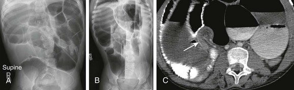
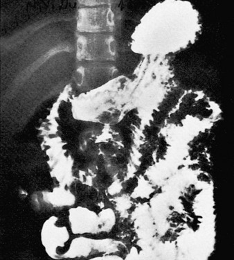
A, Supine radiograph of the abdomen in an 8-year-old boy with a history of multiple abdominal operations and increasing abdominal distension shows dilated, gas-filled loops of bowel. B, Horizontal-beam left lateral decubitus image demonstrates multiple differential air-fluid levels. C, Axial computed tomography image shows a transition zone (arrow) between dilated proximal bowel loops and collapsed distal bowel loops. Adhesions resulting in a small-bowel obstruction were lysed at surgery.
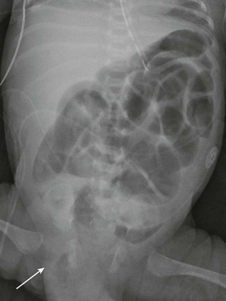
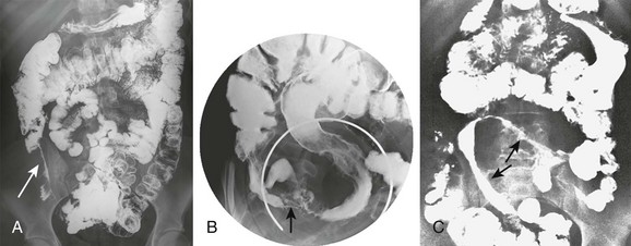
Supine radiograph of the abdomen in a 1-month-old boy, former 26-week gestational age, who presented with a distended abdomen and a right inguinal hernia demonstrates dilated, gas-filled loops of bowel. Bowel gas is seen within the right inguinal hernia (arrow). Obstructive symptoms improved after manual reduction of the hernia.
Treatment
Inflammatory Conditions of the Small Bowel
Overview
Etiology
Clinical Presentation
Imaging
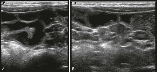
A 21-month-old boy was seen with vomiting and irritability. A, Ultrasonography requested for clinical concern of intussusception reveals distended, fluid-filled loops of bowel that are otherwise normal. B, Additional image in the right lower quadrant reveals multiple enlarged mesenteric nodes, which were hyperemic on color Doppler interrogation (not shown). (Courtesy Marta Hernanz-Schulman MD, Nashville, TN.)
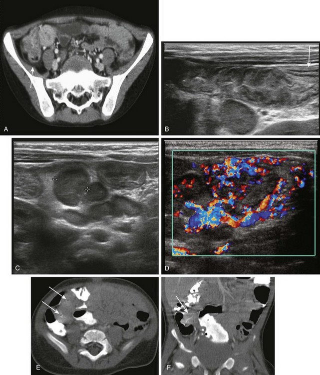
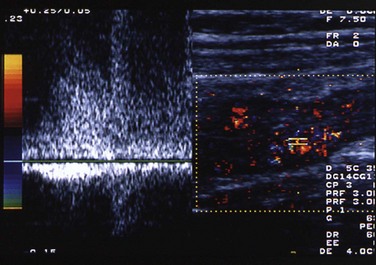
A, Shigella enteritis. Contrast-enhanced computed tomography (CT) reveals concentric thickening and enhancement of the terminal ileum and cecum, along with a small amount of pericecal fluid (arrow). Shigella sonnei was cultured from the stool. B, For a 5-month-old with mucous diarrhea and abdominal pain, ultrasound was requested to assess for suspected intussusception. A transverse sonogram of the right lower quadrant shows a markedly thickened terminal ileum, along with a visible transition to normal bowel (arrow) more proximally. A large lymph node is seen deep to the terminal ileum. C, Transverse image in same infant as shown in B, obtained slightly more cephalad, reveals a large nodal aggregate medial to the ascending colon and anterior to the lower pole of the right kidney (calipers outline one of the nodes). D, Color Doppler image in the same infant shows marked hyperemia in the thick-walled terminal ileum and in the surrounding mesentery and lymph nodes. E, Yersinia enteritis. CT in a 9-month-old boy who had a 10 day history of fever, emesis, and voluminous diarrhea demonstrates pronounced thickening and luminal narrowing of the terminal ileum (arrows). F, Coronal reformat of the CT scan in same patient as shown in E demonstrates similar findings. Stool cultures were positive for Yersinia enterocolitica.
Small intestinal barium examination in a 10-year-old girl with a history of nausea, vomiting, diarrhea, and weight loss. The mucosal folds of the duodenum and the proximal jejunum are markedly thickened. Giardia was identified in the stool.
Treatment
Inflammatory Bowel Disease
Crohn Disease
Etiology
Clinical Presentation
Imaging
A, Frontal abdominal radiograph from small bowel follow-through in a 17-year-old girl with a history of worsening abdominal pain, fever, fatigue, and decreased appetite shows pronounced luminal narrowing of the terminal ileum (arrow) with a “string sign,” which indicates a markedly narrowed segment. Displacement of adjacent bowel loops is evident. B, Spot film of terminal ileum in an adolescent with Crohn disease. Examination reveals extensive involvement and ulceration of the terminal ileum with a “rose thorn” appearance and spasm. Involvement of the cecum is also apparent, and the appendix is incidentally noted at the cecal tip (arrow). C, Small bowel series in a 14-year-old girl reveals a markedly narrowed and rigid loop of mid small bowel (arrows). The upper arrow points to a “string sign.”
Transverse image through the right lower quadrant of an adolescent boy with Crohn disease. Color and spectral Doppler image shows the terminal ileum, which is markedly thickened. Flow to the wall of the terminal ileum is increased, along with venous and high diastolic arterial flow. (Courtesy Marta Hernanz-Schulman MD, Nashville, TN.)
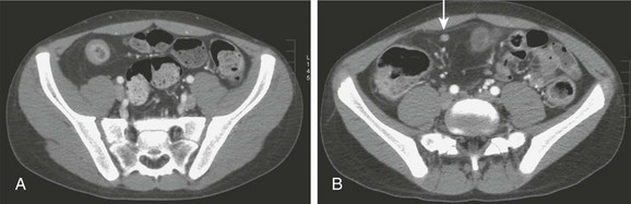
A, Contrast-enhanced computed tomography through the pelvis of an adolescent boy with Crohn disease illustrates the thickened wall of the terminal ileum, hyperemic mucosa, and characteristic circumferential fat deposition, leading to increased separation of bowel loops. B, A section slightly more cephalad in the same patient shows a milder inflammatory change in a more proximal portion of the ileum and again shows fat accumulation, along with a tiny lymph node that is located just deep to the right rectus muscle (arrow).
![]()
Stay updated, free articles. Join our Telegram channel

Full access? Get Clinical Tree


Acquired Lesions of the Small Intestines


