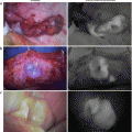
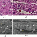
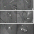
Fig. 27.1
The root of the portal vein (P8ab) feeding segment S8ab was punctured with a 22 G Cattelan needle under ultrasonography (US). After the backflow was confirmed, 1 mL of indocyanine green (ICG) solution (diluted twofold) was injected into the branch of the P8ab under monitoring by US. The indwelling needle 18 G/65 mm then was used to puncture the outside margins of the stained segments under US, and the tip of the outer needle was placed in close proximity of the P8ab
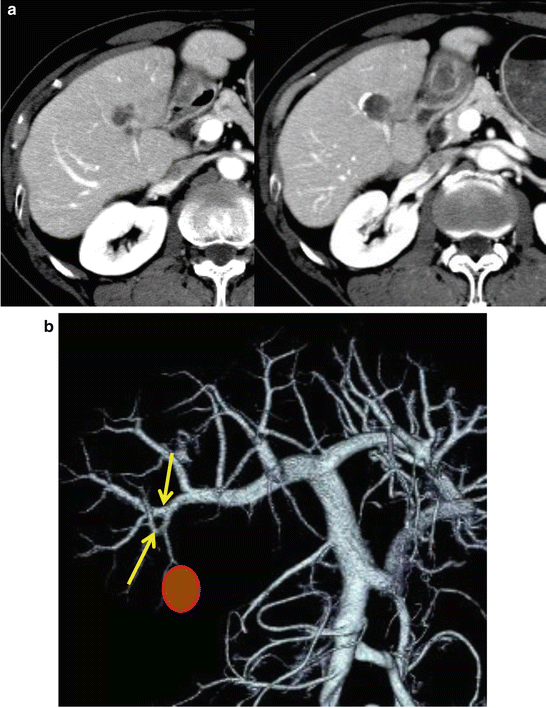
Case: The patient presented a HCC of 3 cm in subsegment S8ab of the liver. Preoperative enhanced CT demonstrated the tumor in the segment fed by the portal vein (P8ab) (Fig. 27.2a). The 3D image reconstructed from a 2D-CT image indicated the root of the P8ab (Fig. 27.2b). Subsequently, laparotomy, cholecystectomy, and intraoperative cholangiography were performed to map the bile duct. The right lobe was removed using the Kocher maneuver to visually expose the boundary between the anterior and posterior sections. The whole liver was examined by intraoperative US to detect the intrahepatic metastasis and to confirm the anatomical structures (i.e., of the branches of the hepatic vein and portal vein). For the occlusion of inflow, the hepatoduodenal ligament was encircled by two elastic tapes. Next, the root of the portal vein (P8ab) feeding segment S8ab was punctured with a 22 G Cattelan needle under US. After backflow was confirmed, 1 mL of diluted (2-fold) ICG solution was injected into the branch of the P8ab while monitored by US. The influx of the ICG solution could be observed by US. If the ICG solution overflowed to the nontargeted portal branches, the injection speed was slowed. Following injection of the ICG solution, the inflow to the liver was successively interrupted by occlusion of the hepatoduodenal ligament to prevent the washout of ICG solution. After dimming of the surgical lights, the surface of the liver was observed with the PDE-neo imaging system. The margin of the S8ab became evident and was marked with an electric scalpel (Fig. 27.3a).
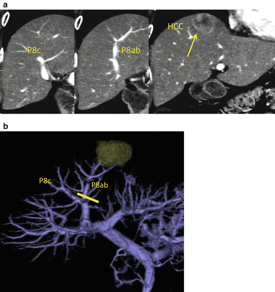
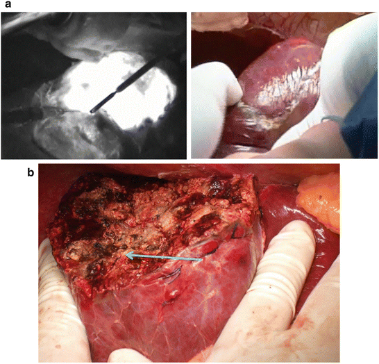

Fig. 27.2
(a) Enhanced computed tomography (CT) demonstrated the tumor in the S8ab subsegment. (b) Three-dimensional (3D)-CT revealed the P8ab was the tumor-bearing portal vein

Fig. 27.3
(a) After the injection of the indocyanine green (ICG) solution, the surface of the liver was observed with the PDE-neo imaging system. The boundary of the S8ab could be distinguished and was marked with an electric scalpel. (b) The parenchyma dissection was completed along the boundary of the S8ab marked with an electric scalpel. The root of P8ab is indicated by the arrow
27.4 Needle-Guiding Technique
Although this method of staining with ICG solution can effectively distinguish segments on the liver surface, it cannot correctly guide to the root of the relevant portal vein and the cut surface. Therefore, to overcome these disadvantages, the indwelling 18 G/65 mm needle is used to puncture the outside of the margin of the stained segments under US, and the tip of the outer needle is placed in close proximity to the P8ab segment (i.e., the needle-guiding technique). This needle is then ligated with a 4-0 silk thread and fixed onto the surface of the liver (Fig. 27.1).
27.5 Hepatectomy
Our standard transection technique of the liver parenchyma is performed using the hook spatula of an ultrasonic harmonic scalpel (Ethicon Endo-Surgery) and a TissueLink DS3.0 monopolar sealer (Classy Med) with soft coagulation mode of VIO300S (ERBE). Vessels are carefully exposed with the hook spatula. Vessels less than 2 mm in diameter are coagulated with the DS3.0 and cut with the hook spatula. Vessels greater than 2 mm in diameter are ligated. Inflow occlusion is applied in an intermittent manner, with 15 min of occlusion alternating with 5 min of reperfusion. During transection of the liver parenchyma, the central venous pressure is maintained below 5 cm H2O to prevent venous hemorrhage.
In the case described above, after the segmentectomy of the S8ab using ICG staining and the needle-guiding technique, the parenchyma was then dissected near the puncture point. After an initial small dissection of the parenchyma, the outer needle was exposed, and the parenchyma was continuously dissected guided by this outer needle. After the cut surface reached the tip of the outer needle, the Glisson of segment S8ab was located and ligated. The parenchyma dissection was completed along the boundary of the S8ab marked with an electric scalpel (Fig. 27.3b).
27.6 ICG Counterstaining
If the puncture of the tumor-bearing portal branch is difficult, a counterstaining identification technique can be used to puncture the adjacent portal branches [12]. Takayama et al. [16] described how under ultrasonic guidance each of the neighboring portal units can be sequentially stained, thus defining the avascular subsegment to be resected as the nonstaining area for the HCC, in which hepatic arterial and portal venous embolizations were already performed.
In our patient, HCC was located in the S6 (Fig. 27.4a, b). This tumor was treated with transarterial chemoembolization (TACE) three times. Due to the cirrhosis present in the noncancerous liver of this patient, the resectable volume of the liver was limited, and because this tumor-bearing portal vein was narrowed, the US-guiding puncture was very difficult to perform. As a result, the two neighboring portal vein branches were punctured, injected with ICG solution, and the surface of the liver was observed using the PDE-neo imaging system. This counterstaining with ICG fluorescent imaging defined the avascular segment to be resected as the nonstaining area and clearly identified the margins between these two areas (Fig. 27.5a, b). The liver fed by the tumor-bearing portal vein was successfully resected.
