The optic pathway, or visual system, conveys sensory information from the retina in the globe to the visual cortex in the occipital lobe. The globe is the primary visual sensory organ responsible for gathering and focusing light and sending electrical and neural signals to the rest of the visual pathway. In the posterior globe, the retina detects incoming light from the environment and converts it into electrical and neural signals that travel along the optic nerve to be processed by the brain for visual perception. Disruptions in the optic pathway and associated clinical signs provide diagnostic information about underlying diseases.
Key points
-
•
The optic pathway, or visual system, conveys sensory information from the retina in the globe to the visual cortex in the occipital lobe.
-
•
Visual sensory information travels via nerve impulses that are triggered by photosensitive chemical reactions in the retina to the optic nerve, tracts, and radiations.
-
•
Disruption of the optic pathway and accompanying clinical signs provide important diagnostic information about underlying diseases.
Abbreviations
| AntCom | anterior commissure |
| Caf | calcarine fissure |
| CaN | caudate nucleus |
| CenB | central bundle |
| CoRa | corona radiata |
| CSF | cerebrospinal fluid |
| DiBandBr | diagonal band of Broca |
| GloPa | globus pallidus |
| IntCap | internal capsule |
| ITG | inferior temporal horn gyrus |
| LGN | lateral geniculate nucleus |
| MTG | middle temporal gyrus |
| OptRad | optic radiations |
| PostB | posterior bundle |
| RGCs | retinal ganglion cells |
| SupLongFasc | superior longitudinal fascicle |
| TH | temporal horn |
Introduction
The following describes the visual pathway in a single hemisphere, proceeding from distal to proximal structures ( Fig. 1 ).
The visual pathway and the corresponding visual field defect resulting from lesions at various locations along the visual pathway.
( Courtesy of Kelly Kage, MFA, CMI.)
Globe
The globe is the primary visual sensory organ responsible for gathering and focusing light and providing electrical and neural signals to the rest of the visual pathway. Light first passes through the globe’s structures, including the cornea, anterior chamber, lens, and posterior chamber, which contains the vitreous ( Fig. 2 ).
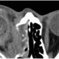
Axial T2-weighted MR imaging of the globe demonstrating the lens ( black arrow ), anterior (∗) and posterior (∗∗) chambers, and the optic nerve ( white arrow ).
The ciliary musculature adjusts the shape of the lens (accommodates) to focus light from various distances upon the retina. The pupil regulates the amount of light that reaches the retina. The visual image is projected backwards and upside-down upon the retina. ,
The ophthalmic artery arises from the internal carotid and supplies most of the blood to the globe. The ophthalmic artery extends into the orbit through the optic canal adjacent to the optic nerve and divides into 2 branches (orbital and extraorbital). The orbital vessels that supply the globe (central retinal artery, muscular artery, and the anterior and posterior ciliary arteries) and the extraorbital vessels supply the other orbital structures (frontal artery, supraorbital artery, lacrimal artery, anterior and posterior ethmoidal arteries, and the nasal artery). ,
Retina
The retina is a multilayered structure composed of photoreceptor, glial, and neuronal cells that lines the posterior segment of the eye, excluding the region occupied by the optic nerve head. The retina detects incoming light from the environment and converts it into electrical and neural signals that travel along the optic nerve to be received by the brain for the perception of a visual image. The retina consists of 10 distinct layers of neurons interconnected by synapses. Within these layers are specialized cells essential for capturing and processing visual information.
Light must first pass through all inner retinal layers before reaching the photoreceptors—rods and cones—which perform the initial phototransduction of visual stimuli and comprise approximately 70% of retinal cells. Retinal ganglion cells (RGCs) serve as the primary output neurons of the retina and include a small subset (approximately 1%–2%) that are intrinsically photosensitive due to their expression of melanopsin, a G-protein-coupled photopigment ( Fig. 3 ). These melanopsin-expressing RGCs contribute to both image-forming and nonimage-forming visual processes, including regulation of the circadian rhythm, melatonin secretion, and the pupillary light reflex. The retina’s constant state of activity makes it one of the most metabolically active tissues in the body, demonstrating the highest oxygen consumption of any tissue.
Anatomy of the retina. The rods, cones, bipolar cells, and ganglion cells are arranged to form a neural pathway. Light strikes the rods (for peripheral and low-light vision) and the cones (for detail and color), which contain photopigments that signal bipolar and ganglion cells. The axons of these cells form the optic nerve, which transmits visual signals to the brain.
( Courtesy of Kelly Kage, MFA, CMI.)
Its dual blood supply is derived from branches of the ophthalmic artery, itself a branch of the internal carotid artery. The outer retinal layers are nourished by diffusion from the choriocapillaris, a capillary layer of the posterior ciliary arteries. In contrast, the inner retinal layers are supplied by the central retinal artery, a principal branch of the ophthalmic artery. Venous drainage occurs via retinal venules, which drain into the central retinal vein. This vein exits the eye in parallel with the central retinal artery through the optic nerve.
Optic nerve
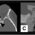
The optic nerve, formed at the level of the retina, transmits afferent light signals to the brain, enabling visual perception. Additionally, the optic nerve serves as the afferent limb and key sensory pathway for the pupillary light reflex and accommodation reflex. The optic nerve is typically subdivided into 4 main segments: the intraocular part (1 mm; the most anterior region of the optic nerve), containing the optic disc; the intraorbital segment (25–30 mm); the intracanalicular (8–10 mm) segment; and the intracranial segment (10–16 mm) ( Figs. 4 and 5 ). Approximately 1.0 to 1.2 million axons of the RGC from the retina gather and form the optic nerve, which later traverses the sclera. The optic nerve head is divided into 4 regions, including the nerve fiber layer, prelaminar region, lamina cribrosa, and retrolaminar region. The lamina cribrosa consists of a multilayered meshwork of collagen fibers that originate from the walls of the scleral canal. After passing through the lamina cribrosa, the optic nerve becomes myelinated by oligodendrocytes and also acquires a dural sheath.
Optic nerve anatomy and segments.
(Yanoff M, Duker JS (eds) Ophthalmology, 2nd edn. St Louis, Mosby, 2004.)
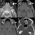
Axial T2-weighted MR imaging demonstrating the 4 mentioned segments of the optic nerve.
( From : Dorigatti Soldatelli M. Seeing is Believing: the Optic Nerve from Anatomy to Pathology. Published online 2017:2371 words. doi:10.1594/ECR2018/C-0112.)
The intraorbital segment of the optic nerve is enclosed by 3 meningeal layers: the dura mater, arachnoid mater, and pia mater. As the optic nerve enters the optic canal, its dural sheath fuses with the periorbita. As it traverses its intracranial course, the optic nerve passes beneath the falciform ligament and is no longer enclosed by a meningeal sheath. Circulation of the cerebrospinal fluid (CSF) around the optic nerve is permitted in the subarachnoid space. The subarachnoid space of the optic nerve is partially connected to the intracranial subarachnoid space at the optic canal and ends at the optic nerve–globe junction. The lamina cribrosa and peripapillary sclera serve as anatomic barriers between the intraocular chamber and the subarachnoid space of the optic nerve, potentially creating a pressure gradient between intraocular fluid and CSF within the optic nerve ( Fig. 6 ). Within the optic canal, the arachnoid mater fuses with the pia mater, indicating a limited continuity and reduced free circulation between the subarachnoid space of the optic nerve and the intracranial subarachnoid compartment in humans. Approximately 1.2 million (with variability in number due to embryogenesis) myelinated nerve fibers exist in each optic nerve. The ophthalmic artery is the major blood supply to the optic nerve, with variation within the 4 segments. In the intraocular region, 2 primary branches emerge at the apex of the orbit, where branches of the central retinal artery and choroid vessels of the posterior ciliary artery (and sometimes through the circle of Zinn-Haller) provide blood supply. Vascularization for the intraorbital, intracanalicular, and intracranial portions is supplied from the superior hypophyseal arteries and branches of the ophthalmic artery, including ciliary arteries.
Anatomy of the optic nerve head. The lamina cribrosa and the surrounding prelaminar (anterior) and retrolaminar (posterior) regions form the optic nerve’s exit point and serve as an anatomic barrier between the intraocular chamber and the optic nerve’s subarachnoid space.
( Courtesy of Kelly Kage, MFA, CMI.)
Optic chiasm
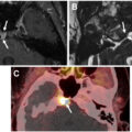
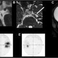
The right and left optic nerves converge at the optic chiasm, which is situated within the subarachnoid space of the suprasellar cistern, approximately 10 mm superior to the sella turcica and the pituitary gland ( Figs. 7 and 8 ). The hypothalamus lies superior to the chiasm, while the infundibulum is positioned posteriorly. Another key anatomic structure in proximity is the cavernous sinus, located inferior to the chiasm and surrounding the pituitary gland. The optic chiasm measures approximately 12 mm in width, 4 mm in thickness, and 8 mm in length, and is inclined at an angle of roughly 45° from the horizontal plane. At the level of the chiasm, a portion of axons from each optic nerve decussate (cross the midline) and merge with the uncrossed fibers from the contralateral optic nerve. A lesion here results in bitemporal hemianopia. The chiasm is so named because it is shaped like the Greek letter chi (χ).
Related posts:
 Orbital Imaging Modalities and Recent Updates
 Imaging of the Globe
 Multidisciplinary Management of Tumors of the Orbit
 Skull Base, Bone, Pituitary—Regions around Orbit that Affect Vision
 Imaging of Central Nervous System Diseases that Affect Vision
 Imaging Findings after Multidisciplinary Treatment for Orbital and Ocular Adnexal Cancers
Stay updated, free articles. Join our Telegram channel

Full access? Get Clinical Tree


