The orbit is a complex anatomic space containing functionally important structures, including the eye, optic nerve, and extraocular muscles. Tumors of the orbit are notably varied in presentation and histologic type and can be challenging to identify, diagnose, and treat secondary to the functional importance of the eye and its surrounding structures. Multidisciplinary expertise from orbital surgeons, radiologists, oncologists, and pathologists is paramount in the timely and accurate management of orbital tumors. Fortunately, with the advancement of surgical approaches, radiation delivery, and targeted medical therapy, patient outcomes and quality of life continue to improve.
Key points
-
•
Orbital tumors present a wide range of pathology in a compact, anatomically complex space.
-
•
The radiologist’s role is paramount in the diagnosis and ongoing management of patients with orbital tumors.
-
•
Treatment of orbital tumors is multifaceted with support from multiple surgical specialists, radiologists, pathologists, and medical oncologists.
Abbreviations
| RMS | rhabdomyosarcoma |
| SCC | squamous cell carcinoma |
Introduction
The orbit is a complex yet compact anatomic space through which pass the vital structures of visual function. Within this conical space, there are several major types of tissues: skeletal and smooth muscle, central and peripheral nervous tissue, vascular structures, bone and connective tissues, and other structures unique to the human eye. Thus, nearly all major types of tumors can occur in the orbit.
Given the importance of each structure in the orbit in terms of visual and ocular function, it is important to consider the impact on patients’ lives when deciding whether and how to intervene for tumors of the orbit. In this article, we will discuss basic principles of diagnosis and management of orbital tumors.
Diagnosis of orbital tumors
Orbital tumors can present insidiously or rapidly. Most benign lesions present in a slower fashion while malignant lesions present more rapidly, although exceptions occur, for example, low-grade lymphomas of the orbit such as marginal zone lymphoma are malignant but usually have a slow onset. Malignant lesions also tend to present with more pain given their rapid growth and increased tendency toward tissue destruction and/or perineural invasion.
Patients may present with proptosis, inflammation around the eye, double vision, or vision loss. These symptoms are corroborated with additional objective findings on physical examination such as change in visual function, optic nerve compromise, and globe displacement. Additional testing with imaging and possibly orbital biopsy will ultimately help with the diagnosis of orbital tumors.
Decision tree for managing orbital tumors
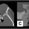
Once an orbital tumor is suspected based on physical examination or imaging findings, it is important to use all the available information to determine what additional steps need to be taken. The first step in deciding how to manage orbital tumors is to decide whether the tumor is presumed to be benign or malignant. This requires input from the history and physical examination along with appropriate imaging studies and, in some cases, a surgical biopsy ( Fig. 1 ).
Decision tree for management of orbital tumors.
The second most important factor to consider is the impact of the tumor on the patient’s visual function. Benign lesions with no impact on visual function may not need any intervention or treatment, whereas those tumors causing objective vision changes, such as loss of vision, double vision, or significant corneal exposure from severe proptosis, may require immediate intervention.
The patient and care team also need to consider the goals of management. Many tumors of the orbit are treated with intent to cure, particularly when removal of the tumor is curative and would not have a significant negative impact on visual and ocular function. In contrast, for metastatic tumors of the orbit, local control in the orbit is usually palliative in nature and treatment strategies are chosen to address the overall disease status. The presence of distant disease involving lymph nodes or other metastatic sites has a significant negative impact on overall survival and thus should factor heavily on how to achieve local and distant control of disease.
For malignant lesions of the orbit without signs of distant metastasis, every effort is made to do eye-sparing surgery, sometimes combined with adjuvant radiation therapy, neoadjuvant immunotherapy, or targeted therapy. In locally advanced cases not responsive to neoadjuvant chemotherapy, immunotherapy, or targeted therapy, sometimes total removal of orbital contents (orbital exenteration) is needed to achieve local control.
Neoadjuvant treatments are aimed at decreasing the orbital tumor size before surgical resection, therefore potentially reducing the need for orbital exenteration. This treatment approach has been particularly useful for tumors originating in the paranasal sinuses with extension into the orbit. Depending on the degree of response to neoadjuvant treatments, sometimes surgical intervention can be avoided entirely. For some orbital tumors, such as lacrimal gland carcinomas or periocular squamous cell carcinoma, adjuvant radiation therapy may be appropriate after eye-sparing surgery. , The recent availability of immune checkpoint inhibitors for periocular squamous cell carcinomas with perineural spread into the orbit and skull base has made it possible to avoid high-dose radiation therapy with its inherent ocular side effects in selected cases.
It is important to discuss the risk of ocular toxicity from treatments for orbital tumors. Surgical interventions for orbital tumors near important structures such as the optic nerve and extraocular muscles are associated with the risk of visual loss from optic nerve damage and diplopia from damage to the extraocular muscles or their innervation. Careful preoperative review of imaging studies of the orbit with a neuroradiologist familiar with orbital anatomy is very helpful to the orbital surgeon in planning the surgical approach and in attempts to avoid damage to intraorbital structures as much as possible. High dose radiation therapy to the orbit is also associated with significant risk of dry eye, optic neuropathy, retinal ischemia, and cataract formation.
Long-term management
The exact nature of the orbital tumor and its treatment will dictate the appropriate long-term follow-up schedule. Serial imaging, particularly MR imaging of orbit, is a very useful radiologic study for this purpose. For example, patients with lacrimal gland carcinoma who have had eye sparing surgery and adjuvant radiation therapy typically have follow up MR imagings every 3 to 4 months for the first 2 years after surgery and then gradually less frequent imaging, but at least once a year for at least 5 years after surgery. In contrast, for patients with low-grade orbital lymphoma such as marginal zone lymphoma, the frequency of imaging studies may be less frequent and limited to a few months after treatments are rendered to assess for response to treatment and then once yearly for 5 years. Patients who have received high-dose radiation therapy will also need ophthalmologic monitoring for potential toxicities involving the ocular surface, tear film insufficiency (dry eye), lens (cataracts), radiation retinopathy, and radiation-induced optic neuropathy. Some medical therapies, for example, immune checkpoint inhibitors, can also cause ophthalmic and orbital complications. Immune checkpoint inhibitors have been associated with the full range of autoimmune-like inflammatory conditions in the eye and orbit: dermatitis, uveitis, myositis, and optic neuritis. , Mitogen-activated protein kinase (MEK) inhibitors are also known to cause vision-altering retinal edema.
Multidisciplinary approach to management of orbital tumors
Most malignant tumors of the orbit and many of the benign tumors require multidisciplinary management. The neuroradiologist plays a critical role in providing input to the orbital surgeon regarding the likely diagnosis or a list of possible diagnoses. The size and extent of the orbital mass, proximity to the extraocular muscles and optic nerve, involvement of the bony walls of orbit, extension toward the superior orbital fissures, presence of obvious perineural spread, and in some cases, involvement of paranasal sinuses, the nasal cavity, and skull base are all important details to address in the initial radiology report.
Once an orbital mass is biopsied or surgically resected by the ophthalmic plastic surgeon, the expertise of a pathologist familiar with orbital pathology is important in making the correct diagnosis. After this step, various treatment options can be discussed at a multidisciplinary tumor board, ideally with input from medical oncologists and radiation oncologists. Finally, it may be important to engage other supportive services such as prosthodontics, social work, physical and occupational therapy, and palliative and hospice care in advanced cases.
Surgical treatment of an orbital mass may also require multiple surgeons from various complementary disciplines. If the orbital mass is contained in the orbital cavity, the orbital/oculoplastic surgeon can often resect the lesion in its entirety or perform an orbital exenteration if eye-sparing surgery is not possible. However, for lesions that extend to the nasal cavity and paranasal sinuses, the base of skull, or extend intracranially, head and neck surgery, neurosurgery, skull base surgery, and plastic surgery expertise may be called upon.
Management of suspected benign tumors
Several benign entities can arise in the orbit, which may or may not cause symptoms or clinical findings. Many masses within the orbit may be found incidentally while undergoing radiographic evaluation for unrelated problems, such as headaches or as part of a trauma evaluation. For example, cavernous venous malformations (cavernous hemangiomas) and other vascular malformations, Schwannomas, and meningiomas are sometimes found in completely asymptomatic patients incidentally.
Serial imaging using MR imaging or CT scans can be helpful in monitoring benign orbital masses that are being observed. For benign asymptomatic lesions such as cavernous venous malformation, asymptomatic schwannoma, or meningioma, a typical frequency for repeating orbital imaging may be every 6 months initially, followed by annual imaging if stability is noted. If growth is noted over time, a closer interval may be warranted. The choice of which imaging to perform should consider which images demonstrate the salient features of the lesion with consideration to the recurring cost to the patient, exposure to contrast and/or radiation, and any other patient factors which may limit the imaging choices such as presence of pacemakers or other devices that may be a relative contraindication to MR imaging.
Surgery is the most common treatment of benign orbital tumors that cause symptoms such as visual loss, visual field deficit, gaze-evoked amaurosis, significant proptosis, corneal exposure, diplopia especially in primary gaze, and choroidal and globe distortion. The specific surgical approaches to the orbit will be discussed in more detail in the next section. The most common benign orbital lesions that are addressed with gross total resection with an intact capsule or pseudocapsule are cavernous venous malformation, schwannomas, sphenoid wing meningiomas, dermoid cysts, and pleomorphic adenoma of the lacrimal gland.
Some benign neuronal lesions of the orbit, such as optic nerve sheath meningiomas, may be considered for radiation therapy. Radiation has also been used for optic nerve gliomas, particularly when progression is noted to negatively affect the eye or threaten encroachment into the optic chiasm or nerve from the opposite eye. Radiation can also be considered for recurrent cases of orbital inflammatory syndrome or immunoglobulin G4 (IgG4) disease.
More recently, targeted medical therapies may be available for some lesions; for example, neurofibromas involving the orbit, while considered benign, can have negative effects on vision and appearance. Sparing the patient from radical surgery or radiation therapy, particularly in children, motivated the development and use of selumetinib with good effect. For venolymphatic malformations, sildenafil given systemically and/or sclerosing agents delivered through interventional radiology techniques have been used successfully. , Systemic and topical beta blockers have been successfully used for the treatment of periorbital capillary hemangiomas. Some of these vascular lesions can also be successfully treated with procedures such as embolization for high-flow vascular lesions.
Management of suspected malignant tumors
Certain features of orbital tumors will lead the suspicion for a malignant process. Orbital lesions that are rapidly progressive and/or painful warrant careful investigation for possible malignancy; pain can be the result of perineural invasion or tissue destruction not typical of more benign entities. Obviously, when there is a clinical history of oncologic disease, particularly if recent, the possibility of metastatic disease to the orbit must be considered. Metastatic disease traditionally involves the extraocular muscles or other soft tissue or orbit, and the bony walls of the orbit, such as the greater wing of the sphenoid bone. Patients with epistaxis or bloody tears may harbor malignant lesions involving the sinuses, nasal cavity, or nasolacrimal system.
When a malignant orbital tumor is suspected, getting an accurate diagnosis is essential and often requires obtaining tissue for pathologic analysis. However, there are scenarios when an incisional biopsy is more appropriate and others when complete excisional biopsy is the appropriate form of obtaining tissue diagnosis. For example, for a lesion suspected to be lymphoma or orbital inflammatory disease, an incisional biopsy is appropriate. On the other hand, in the case of presumed pleomorphic adenoma of the lacrimal gland, there is a concern that incisional biopsy or incomplete excision can lead to multifocal benign recurrences that are debilitating for the eye and its function over time, or, more rarely, they can undergo transformation into frank carcinoma. , Thus, preoperative review of imaging studies and discussion with the neuroradiologist on the care team can be extremely helpful in narrowing down the differential before deciding on an excisional versus incision biopsy for a newly diagnosed lacrimal gland mass.
For metastatic lesions in the orbit, if there is a previous history of cancer and the patient is known to have multiple other sites of metastasis throughout the body, it may not be necessary to do an orbital biopsy. But in the case of a suspected oligometastatic lesion to the orbit or in patients with a history of multiple previous cancer types, it may be necessary to do an incisional biopsy to establish a diagnosis; gross total resection of the mass can be considered if there is minimal ocular morbidity. Surgical debulking of an orbital metastasis may be considered in some cases to relieve or improve symptoms of mass effect. Otherwise, most cases of orbital metastasis are treated either with systemic chemotherapy, targeted therapy, or immunotherapy or with palliative low-to moderate-dose radiation therapy. It should also be noted that when orbital metastases are discovered, the disease is often at an advanced stage, and the merits of curative versus palliative treatment options should be carefully considered.
For primary malignant orbital lesions, surgical resection is the mainstay of treatment of most cases. The main consideration is whether eye-sparing surgery can be done versus orbital exenteration. For lacrimal gland carcinomas, the field has moved significantly toward eye sparing approaches. Approximately 75% of patients with lacrimal gland adenoid cystic carcinoma have eye-sparing surgery followed by adjuvant radiation therapy. The rest have orbital exenteration. In the era of eye-sparing treatments for patients with lacrimal gland carcinoma, it is particularly important for neuroradiologists to closely collaborate with the orbital surgeon in the surgical planning stage as well as during the follow-up period of several years. Serial imaging is important to rule out local recurrence for at least 5 years after surgery in patients with lacrimal gland carcinoma.
For the most common pediatric orbital tumor, orbital rhabdomyosarcoma, the initial management is surgical debulking and a biopsy to establish the diagnosis as quickly as possible. Treatments consist of chemotherapy and radiation therapy to the orbit; most patients avoid orbital exenteration and more than 90% of patients with pediatric orbital RMS are cured of their disease with modern multidisciplinary treatments.
Medical management of malignant orbital disease can be in the neoadjuvant or adjuvant setting. Neoadjuvant chemotherapy or immunotherapy using immune checkpoint inhibitors can be used to decrease the size of tumor before surgical removal. Immune checkpoint inhibitors are gaining increasing popularity for treatment of locally advanced squamous cell carcinoma (SCC) of the orbit and periocular region and especially for patients with cutaneous SCC of the head and neck area with perineural spread into the orbit and skull base. Immune checkpoint inhibitor (ICI)’s also have efficacy for locally advanced or metastatic conjunctival melanoma and periocular cutaneous melanomas as well as for locally advanced basal cell carcinoma (BCC) with orbital invasion that is refractory to treatment with sonic hedgehog inhibitors. Availability of targeted drugs and immune checkpoint inhibitors in the last decade have led to a decrease in the number of patients who require orbital exenteration. A summary of available therapies specific to tumors commonly involving the orbit and periocular tissues is shown in Table 1 .
Table 1
Targeted and immunotherapy options for orbital tumors
| Disease | Mechanism of Action | Examples |
|---|---|---|
| Basal cell carcinoma | Sonic hedgehog inhibitor | Vismodegib and sonidegib |
| Squamous carcinoma | PD-1 inhibitor | Cemiplimab, pembrolizumab, and nivolumab |
| EGFR inhibitor | Cetuximab | |
| Melanoma | BRAF inhibitor/MEK inhibitor | Vemurafenib |
| PD-1 inhibitor | Pembrolizumab and nivolumab | |
| CTLA4 inhibitor | Ipilimumab | |
| MAP kinase inhibitor | Dabrafenib | |
| Merkel cell carcinoma | PD-L-1 inhibitor | Selumetinib |
| PD-1 inhibitor | Pembrolizumab and nivolumab | |
| NF | MEK Inhibitor | Selumetinib |
| Pediatric glioma | RAF kinase inhibitor | Tovorafenib |
| Histiocytic disorders | BRAF inhibitor/MEK inhibitor | Vemurafenib |
| Lymphoma | Anti-CD20 | Rituximab |
| Uveal melanoma | T-Cell mobilization | Tebentafusp |
| PKC inhibitor | Darovasertib |
Related posts:
Stay updated, free articles. Join our Telegram channel

Full access? Get Clinical Tree


