Arteries of the Head and Neck
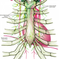
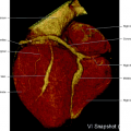
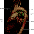
The arterial vascularization of the head and neck originates from the three main arteries at the aortic arch (Fig. 2.1). In two thirds of the population, the brachiocephalic trunk is the first vessel that originates from the aortic arch, the left carotid artery is the second, and the left subclavian artery is the third. The right common carotid begins at the bifurcation of the brachiocephalic trunk and the right vertebral artery originates from the right subclavian artery, also a branch of the brachiocephalic trunk. The left common carotid artery arises directly from the aortic arch, whereas the left vertebral artery originates from the left subclavian artery (Fig. 2.2).
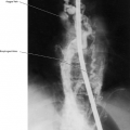
In one third of individuals, three most common variations may be encountered: (1) the left common carotid artery either has a common origin with the brachiocephalic trunk or originates from the proximal portion of this trunk (most common variation) (Fig. 2.3); (2) the left vertebral artery originates directly from the aortic arch between the left common carotid artery and left subclavian artery (Fig. 2.4); (3) there is aberrant origin of the right subclavian artery from the aortic arch distal to the left subclavian artery, usually retroesophageal when crossing the mediastinum (lusory artery, present in 0.2% to 2.5% of the population) (Fig. 2.5).
Common Carotid Artery
The common carotid artery has thoracic and cervical portions. It is enclosed within the carotid sheath, together with the vagus nerve and the jugular vein. The common carotid arteries ascend from the arch of the aorta, in front of the trachea, to the cervical portion, where they incline laterally to both sides of the trachea (Figs. 2.1, 2.2, 2.6). The left common carotid artery is usually longer than the right common carotid artery, and in individuals with short necks, the level of the bifurcation of both common carotids is higher.
The common carotid arteries usually have no branches, either in the thoracic portion or the cervical part, but may give origin to the vertebral, the superior thyroid or its laryngeal branch, the ascending pharyngeal, the inferior thyroid, or the occipital artery. At the level of the upper border of the thyroid cartilage, the common carotid arteries bifurcate into the external and internal carotid arteries (Figs. 2.7, 2.8). At the division, the vessel dilates and is called the carotid sinus, which usually involves only the origin of the internal carotid artery. The carotid sinus contains a large number of sensory nerve endings, from the glossopharyngeal nerve, acting as a baroreceptor mechanism that exercises control over the intracranial pressure. The carotid body lies behind the level of the bifurcation of the common carotid artery and has a chemoreceptor function.
External Carotid Artery
The external carotid artery arises medial and anterior to the internal carotid artery (Figs. 2.9, 2.10). Occasionally it may arise lateral to the internal carotid artery, particularly in older individuals.
Anterior Branches
Superior thyroid artery
Lingual artery
Facial artery
Posterior Branches
Ascending pharyngeal artery
Occipital artery
Posterior auricular
Terminal branches
Superficial temporal artery
Internal maxillary artery
Superior Thyroid Artery
This artery arises from the external carotid artery, as the first anterior branch, just below the level of the greater cornu of the hyoid bone, and divides into terminal branches at the apex of the lobe of the thyroid gland. It may arise from the common carotid artery.
Branches
Anterior branch or superior marginal arcade (anastomoses with the opposite artery through the isthmus)
Posterior branch or posterior glandular arcade (anastomoses with the inferior thyroid artery)
Lateral branch or lateral glandular arcade (not constant)
Hyoid branch (anastomoses with the thyrolaryngeal system inferiorly with the linguofacial system superiorly)
Sternocleidomastoid artery
Superior laryngeal artery (anastomoses with the opposite artery and the inferior laryngeal artery)
Cricothyroid artery (anastomose with the opposite artery)
Lingual Artery (Figs 2.12, 2.13)
This artery is the second branch of the external carotid artery and main feeder of the tongue muscles of the floor of the mouth and the sublingual gland. It arises from the anteromedial aspect of the proximal external carotid artery, between the origin of the superior thyroid artery and the facial artery. Occasionally it may have a common origin with the facial artery constituting the linguofacial trunk (Fig. 2.7). This artery runs obliquely upward and medially, curving downward and forward and forming a loop. It runs horizontally forward and finally ascends sharply in the cranial direction, coursing under the surface of the tongue as far as its tip.
The lingual artery may be divided in three parts. The first part is in the carotid triangle. The second part of the lingual artery traverses the upper border of the hyoid bone, deep to the hyoglossal and the lower part of the submandibular gland. The hyoglossal separates the artery from the hypoglossal nerve and its vena comitans. The third part of the artery is also called arteria profunda linguae. It runs close to the tongue and is accompanied by the lingual nerve. At the tip of the tongue it anastomoses with the contralateral artery.
Branches
Suprahyoid branch (small, anastomoses with the contralateral artery)
Dorsal artery of the tongue (largest branch supplying the tongue)
Sublingual artery (supplies the sublingual gland and neighboring muscles and mucous membrane of the mouth and gums. Anastomoses with the submental artery arising from the facial artery. A medial mandibular branch supplies the anterolateral surface of the body of the mandible. Depending on the hemodynamic balance of the region, the lingual artery, through its anastomotic branches, can take over the supply of the gland and the mandible, and occasionally part of the submental territory [Figs. 2.13, 2.14]).
Originates from the anterior aspect of the external carotid artery, as the third branch, still in the carotid triangle, just above the lingual artery and the greater cornu of the hyoid bone. Runs medial to the ramus of the mandible causing a groove on the posterior border of the submandibular gland. It turns downward and forward, reaching the lower border of the mandible and becoming superficial and subcutaneous. At this point, the main facial trunk can have two different courses, a more posterolateral or jugal course, or a more anteromedial or labial course (Fig. 2.15). The facial artery turns cranially to the side of the nose, ending at the medial palpebral commissure, supplying the lachrymal sac and anastomosing with the dorsal nasal branch of the ophthalmic artery.
The facial artery supplies the muscles and tissues of the face, the submandibular gland, the tonsil, and the soft palate. The branches may be separated in cervical and facial groups. There are abundant anastomoses of the facial artery, not only with the contralateral branches of the vessel at the opposite side but also in the neck (with the sublingual branch of the lingual artery and with the palatine branch of the maxillary) and in the face (with the mental branch of the inferior alveolar artery, the transverse facial branch of the superficial temporal artery, the infraorbital branch of the maxillary, and the dorsal nasal branch of the ophthalmic artery). The territory vascularized by the facial artery is in hemodynamic equilibrium with the adjacent arteries that may be part of the facial artery territory (Figs. 2.13, 2.14, 2.16, 2.17).
Branches
Ascending palatine artery (arises close to the origin of the facial artery and runs upward at the side of the pharynx, in the pillar area) Divides in two branches: (1) to the muscle levator veli palatine and soft palate (Fig. 2.19), where it anastomoses with a branch of the descending palatine artery; (2) the other branch penetrates the superior constrictor and supplies the tonsils and the auditory tube. (Anastomoses are with the tonsillar, accessory meningeal, and ascending pharyngeal artery and with its counterpart on the other side.) The ascending palatine artery or artery of the soft palate may arise directly from the external carotid artery (Fig. 2.18), from the ascending pharyngeal artery (Fig. 2.19), or from the accessory meningeal artery.
Tonsillar artery (supplies the tonsil and root of the tongue)
Glandular branches (Three or four branches supplying the submandibular salivary gland, lymph nodes, and neighboring muscles and skin) (Fig. 2.15).
Submental artery (Largest cervical branch. Supplies the musculocutaneous region of the mandible and chin, and anastomoses with the sublingual branch of the lingual artery and mylohyoid of the inferior alveolar artery. It divides in superficial and deep branches.) (Figs. 2.13, 2.14). The submental artery replaces sometimes the entire facial trunk, when it is hypoplastic.
Inferior labial artery (arises at the angle of the mouth and extends near the edge of the lower lip between the muscle and mucous membrane. Anastomoses with the contralateral artery and the mental branch of the submental artery).
Superior labial artery (courses along the edge of the upper lip between the muscle and mucous membrane). Anastomoses with the contralateral artery. It gives off a septal branch to the lower and frontal part of the nasal septum, and an alar branch to the ala of the nose.
Lateral nasal branch (also called the angular artery, this vessel ascends along the side of the nose. It supplies the alar artery and the nasal arcade at the dorsum of the nose, anastomosing with the contralateral artery, the septal and alar branches of the superior labial artery, and with the dorsal nasal ramus of the ophthalmic artery and the infraorbital branch of the maxillary artery)
Inferior masseteric artery (arises from the facial artery after it has passed under the mandible. It anastomoses with the middle and superior masseteric arteries)
Jugal trunk. It includes two different functional units.
The buccomasseteric system or buccal branch. It anastomoses with the facial artery and with the maxillary artery in the upper part of the pterygopalatine fossa. It supplies the deep muscle–mucosal structures and constitutes the preferential collateral pathway between both systems.
The posterior jugal artery, which pursues a superficial course connecting the lower border of the mandible with the external orifice of the infraorbital canal; here it anastomoses with the infraorbital artery, the superior alveolar artery, and the anterior and middle jugal branches.
Middle mental artery (arises midway up the lateral surface of the body of the mandible)
Anterior jugal artery (supplies the anterior part of the jugal area and anastomoses with the posterior and middle jugal arteries)
Internal Maxillary Artery (Fig. 2.11)
This is the larger terminal branch of the external carotid artery; it arises behind the neck of the mandible and it is proximately embedded in the parotid gland, subsequently passes close to the lower head of the lateral pterygoid muscle, and distally enters to the deep of the pterygopalatine fossa between the two heads of that muscle. It may be divided into three segments: mandibular, pterygoid, and pterygopalatine.
Mandibular Segment (Behind the neck of the mandible)
Branches of the Mandibular Segment
Deep auricular artery (small, may be a branch of the anterior tympanic artery. Supplies the outer aspect of the tympanic membrane and temporomandibular joint)
Anterior tympanic artery (supplies the medial aspect of the tympanic membrane and anastomoses with the posterior tympanic branch of the stylomastoid artery)
Middle meningeal artery (largest meningeal artery) (Figs. 2.20, 2.21, 2.22). This artery enters the cranial cavity through the foramen spinosum of the sphenoid bone. It runs forward and laterally in a temporal bone groove and vascularizes large areas of the supratentorial meninges and anastomoses with other meningeal branches and with the ophthalmic artery. It may give origin to the ophthalmic artery or to its glandular and muscular branches (meningolacrimal artery) (Fig. 2.23).
Frontal branch (anterior)
Parieto-occipital branch (posterior)
Petrosquamosal trunk
Accessory meningeal artery (Fig. 2.22) It may be a branch of the maxillary artery (Fig. 2.23) or the middle meningeal artery (Fig. 2.20). Enters the cranium through the foramen ovale. It has an extracranial branch that goes to the cavum at the pharyngotympanic tube level and another intracranial branch anastomosing with branches of the internal carotid, ophthalmic artery, and middle meningeal artery.
Inferior alveolar (dental) artery (Figs 2.16, 2.20). (Arises from the proximal portion of the internal maxillary artery and follows a descending direction. It enters the mandibular canal at the internal surface of the mandible together with the nerve and inferior alveolar vein. Anastomoses with the
submental artery, branch of the facial artery, and originates the mylohyoid branch.)
Pterygoid Segment (Superficial or Deep to the Lateral Pterygoid Muscle in the Temporal Fossa)
Branches of the Pterygoid Segment
Deep temporal branches (Figs. 2.20, 2.24) (Anterior, middle, and posterior. They supply the temporal muscle. These vessels distinguish themselves by the straightness of their course and by the fact that their course is not altered at the base of the skull. The anterior branch anastomoses with the lachrymal artery, through the zygomatic and sphenoid bones.)
Pterygoid branches (supply the pterygoid muscle)
Masseteric arteries (Figs. 2.20, 2.16) (supply the masseter, a muscle of mastication. This muscle is supplied by four groups of vessels: superior, middle, inferior, and deep masseteric arteries.)
Buccal artery (Fig. 2.20) (runs along the buccal nerve, to the buccinator muscle, and anastomoses with branches of the facial and infraorbital arteries. This branch constitutes the most important connection between the maxillary and facial systems. It arises from the distal part of the maxillary artery and descends vertically posterior to the maxillary tuberosity)
Pterygopalatine Segment
This segment enters the pterygopalatine fossa and terminates by dividing into several branches denominated according to the direction with which they exit from the fossa.
Branches of the pterygopalatine segment (Figs. 2.16, 2.20)
Superior alveolar (dental) artery (originates as the maxillary artery and enters the pterygopalatine fossa. Gives several branches: some to the alveolar canals and others to the alveolar process to supply the gums.)
Infraorbital artery (Fig. 2.20). (This artery is the most anterior branch of the maxillary artery. It defines the superior boundary of the maxillary sinus and corresponds to the most inferior part of the orbit. It enters the inferior orbital fissure and emerges with the infraorbital nerve on the face through the infraorbital foramen. On the face, there are anastomoses with terminal branches of the facial artery, a dorsal nasal branch of the ophthalmic artery, and transverse facial and buccal arteries.)
Greater palatine artery (Fig. 2.20) (This artery runs through the greater palatine canal and gives off two or three lesser palatine arteries to the soft palate and tonsil. It anastomoses with the ascending palatine artery and branches of the sphenopalatine artery.)
Pharyngeal branch (It is very small and distributes to the mucosa of the nose, pharynx, sphenoidal air sinus, and auditory tube.)
Artery of the pterygoid canal (is a branch of the greater palatine artery and feeder of the upper pharynx, the auditory tube, and the tympanic cavity)
Sphenopalatine artery (Fig. 2.20) (The real terminal part of the maxillary artery. Passes into the nose at the posterior part of the superior meatus. The branches are the posterior lateral nasal branches, with anastomoses with the ethmoidal arteries and the nasal branches of the greater palatine artery. The sphenopalatine artery ends on the nasal septum as the posterior septal branches and anastomoses with the ethmoidal arteries, the terminal ascending branch of the greater palatine artery, and the septal branch of the superior labial artery.)
This artery arises close to the origin of the external carotid and ascends vertically between the internal carotid and the side of the pharynx to the base of the skull. It has two divisions, one is anterior and the other posterior, also called neuromeningea.
The anterior division gives origin to pharyngeal branches (superior, middle, and inferior) (Fig. 2.19) and to the inferior tympanic artery (Fig. 2.25), which may be a single independent branch. The posterior division gives origin, distally, to a jugular branch (enters the jugular foramen and feeds the IX, X, and XI nerves) and a branch called hypoglossal nerve branch (enters the hypoglossal canal and feeds the hypoglossal nerve, reaching the meninges of the posterior fossa) (Fig. 2.19). The hypoglossal branch may give origin to the odontoid arcade, which vascularizes the meninges close to the odontoid process.
The two branches of the posterior division anastomose with the clival branches (Fig. 2.22) of the meningohypophyseal trunk from the internal carotid artery (Fig. 2.26). Another branch of the posterior division is the musculospinal artery, oriented downward and posteriorly, supplying the XI nerve and the superior sympathetic ganglion. The ascending pharyngeal artery may arise from the external carotid artery, from a common trunk with the occipital artery (Fig. 2.27), or from the internal carotid artery (Fig. 2.28). It has anastomoses with the vertebral artery at the second and
third cervical levels (Figs. 2.19, 2.26) and with the internal carotid artery (Fig. 2.25).
third cervical levels (Figs. 2.19, 2.26) and with the internal carotid artery (Fig. 2.25).
The occipital artery is a posterior branch of the external carotid artery (Fig. 2.7). It arises at the level of the facial artery in the opposite direction. The artery runs backward and upward, crossing the internal carotid artery; the internal jugular vein: and the hypoglossal, vagus, and accessory nerves. The distal artery reaches the space between the transverse process of the atlas and mastoid process of the temporal bone. It then runs in the occipital groove on the temporal bone, where it is medial to the mastoid process and the attachment of the sternocleidomastoid and other muscles. Distally it turns upward and divides into several smaller branches.
Branches
Sternocleidomastoid branches (Lower and upper branches supply the muscle.)
Mastoid branch (Usually small and sometimes absent; enters through the mastoid foramen to feed the mastoid air cells and dura mater at the level of the cerebellopontine angle. Anastomoses with the middle meningeal artery.)
Auricular branch (supplies the back of the auricle and anastomoses with the posterior auricular artery)
Muscular branches. There are several unnamed muscular branches; the most important of them follows a pattern determined by the intervertebral spaces. For each posterior space, a parasagittal branch gives rise to a posterior anastomotic radicular branch and a lateral branch. There are anastomoses with the vertebral artery in the first three cervical intervertebral spaces (Fig. 2.29).
Meningeal branches. Two branches supply the meninges of the posterior cranial fossa: (1) the artery of the falx cerebelli (Fig. 2.27)—this artery arises from the anastomoses in the first cervical space—and (2) the mastoid branch.
Posterior Auricular Artery
This is a small artery, arising directly from the posterior aspect of the external carotid artery (Fig. 2.7). It supplies muscles and the parotid gland, and it has three main branches. It has a hemodynamic equilibrium with the occipital and temporal superficial arteries (Fig. 2.24)
Branches
Stylomastoid artery (Fig. 2.31). This artery enters the stylomastoid foramen, and supplies the tympanic cavity, the mastoid antrum, the mastoid air cells, and the semicircular canals. In youngsters, the posterior tympanic artery forms a vascular circle surrounding the tympanic membrane.
Auricular branch (supplies the auricularis posterior)
Occipital branch (anastomoses with the occipital artery)
Superficial Temporal Artery (Fig. 2.9)
This is one of the terminal branches of the external carotid artery. It arises close to the parotid gland, behind the neck of the mandible, and has an anterior and a posterior branch (Fig. 2.24). This artery supplies the parotid gland, the temporomandibular joint, the masseter, the auricula, and the skin and scalp.
Branches
Transverse facial artery (Fig. 2.31). This vessel arises from the parent artery inside the parotid gland. It divides into numerous branches that extend to the parotid gland and duct, the masseter, and the skin. Anastomoses with the facial, masseteric, buccal, lachrymal, and infraorbital arteries.
Anterior auricular branch (supplies the lobule, the anterior part of the auricle, and the external acoustic meatus)
Zygomatico-orbital artery (Occasionally it is a branch of the middle temporal artery and supplies the orbicular muscles and anastomoses with branches of the ophthalmic artery, and lachrymal and palpebral arteries)
Middle temporal artery (anastomoses with the deep temporal branches of the maxillary artery)
Frontal (anterior) branch (runs upward and forward over the frontal bone. It is tortuous and anastomoses with the contralateral artery)
Parietal (posterior) branch (runs upward and backward on the side of the head. Anastomoses with the posterior auricular and occipital arteries)
Internal Carotid Artery
The internal carotid artery originates from the bifurcation of the common carotid artery, in general at the level of the fourth cervical vertebra, or the superior border of the thyroid cartilage, in adults. It usually lies posterior and lateral to the external carotid artery (Figs. 2.1, 2.6). It may, however, have a course anterior and medial to the external carotid artery. In about half of the cases, the internal carotid artery presents a fusiform dilation near the origin called the carotid sinus. Beyond the carotid sinus the caliber of the internal carotid artery is uniform (Fig. 2.30).
The internal carotid artery has three main segments: cervical, petrous, and intracranial segments.
Cervical Segment
At the cervical segment, the internal carotid artery is almost vertical, from the origin to the carotid canal at the base of the skull. It is closely connected to the jugular vein and to the vagus nerve, which lies behind and between these two vessels, forming a neurovascular bundle. It has two parts: one lower part localized at the sternocleidomastoid region and an upper part localized at the retrostyloid region. Elongation, loops, and tortuosity are common in older patients and are accentuated by flexion of the neck and straightened by extension of the neck.
Petrous Segment
There is a vertical and a horizontal portion of the petrous segment. The vertical portion passes inside the petrous bone for about 1 cm and then turns anteriorly and medially. The horizontal portion passes forward and medially within the petrous bone to emerge near the apex of the bone.
Intracranial Segment
The intracranial portion of the internal carotid artery may be divided into three segments: the precavernous segment, the cavernous segment, and the supraclinoid segments.
The precavernous segment courses upward, forward, and medially from the apex of the petrous temporal bone to the point where it is related to the lower and posterior aspects of the sella turcica, before entering the cavernous sinus. It is also called ganglionar segment because it is in contact with the gasserian ganglion, located laterally to the internal carotid.
The cavernous segment lies within the cavernous sinus and ascends a short distance lateral to the lower and posterior aspects of the sella. At the carotid sulcus, this segment passes anteriorly on the lower and lateral aspects of the sella, and then curves upward, medial to the anterior clinoid process. Within the cavernous sinus the sixth (VI) nerve lies on the lateral aspect of the artery. The third (III), the fourth (IV), the ophthalmic (V), and the maxillary nerves are closely related to the lateral wall of the cavernous sinus.
The supraclinoid segment courses upward, after crossing the dura, medial to the anterior clinoid process, posteriorly, and laterally to its point of bifurcation. The optic nerve lies medial to the lower part of this segment (Fig. 2.31).
Branches
Mandibular artery
Caroticotympanic branch
Meningohypophyseal trunk
Basal tentorial branch
Inferior hypophyseal artery
Clivus branches
Inferolateral trunk
Marginal tentorial branch
Branches to the gasserian ganglion; IV, V, and VI nerves; and to the wall of cavernous sinus. Branches to the orbit
Superior hypophyseal branches
Ophthalmic artery
Posterior communicating artery
Anterior choroidal artery
Anterior cerebral artery (terminal branch of internal carotid artery)
Middle cerebral artery (terminal branch of internal carotid artery)
Mandibular Artery
It arises from the petrous segment, either in the foramen lacerum or in the horizontal portion at the carotid canal.
Caroticotympanic Branch
Arises from the posterior, distal, vertical petrous segment of the internal carotid artery. This small branch penetrates the tympanic cavity and anastomoses with the inferior tympanic branch of the ascending pharyngeal artery (Fig. 2.25), the anterior tympanic branch of the maxillary artery, and the stylomastoid artery.
The branches of the meningohypophyseal trunk arise from the posterior aspect of the internal carotid artery. The tentorial branch, inferior hypophyseal artery, and clival branches arise from the dorsal main stem.
Branches
Basal tentorial branch (Fig. 2.32). Enters the tentorium anterior to the apex of the petrous bone and continues in the tentorium near the tentorium attachment to the petrous bone (tentorial basal), supplying the adjacent tentorium, or at the free edge of the tentorium (Marginal of Tentorium).
Clivus branches (Fig. 2.31). Supply the dura of the dorsum sellae and clivus. Anastomose with the contralateral corresponding arteries.
Inferior hypophyseal artery (Figs. 2.31,2.32,2.33). Supplies the posterior lobe of the gland.
Inferolateral Trunk (Fig. 2.31)
The inferolateral trunk arises more anteriorly from the lateral and inferior aspects of the internal carotid artery. It passes downward and laterally over the lateral aspect of the sixth nerve and further downward through lateral or under the fifth nerve. It also gives branches to the gasserian ganglion; the fourth, fifth, and sixth nerves; and the wall of the cavernous sinus. The main vessel supplies the dura in the floor of the middle fossa and anastomoses with branches of the middle meningeal artery and accessory meningeal artery, whereas other small branches go anteriorly and through the superior orbital fissure or directly through the greater wing of the sphenoid to the orbit, where they anastomose with branches of the Ophthalmic Artery.
Superior Hypophyseal Branches
These are branches of the internal carotid artery at the level of the supraclinoid segment or Posterior Communicating Artery. They supply the pituitary stalk and the anterior lobe of the pituitary gland.
Ophthalmic Artery
The ophthalmic artery (Fig. 2.17) arises immediately above the superior limit of the cavernous sinus. It is the first major branch of the internal carotid artery. In 83% of the cases the origin of the ophthalmic artery is in the subdural space at the level where the internal carotid artery exits the cavernous sinus, penetrating the dura (Fig. 2.6). In 6.5% of cases it may be slightly distal to this site by 1 mm or so. In 7.5% of the cases it may be extradural, arising from the intracavernous portion of the internal carotid artery (Fig. 2.33). In 2% of the cases it arises from the internal carotid artery, at the level it penetrates the dura.
Other Origins and Anastomoses of the Ophthalmic Artery
The anomalous origins of the ophthalmic artery depend on the embryologic development established by this artery with the adjacent vessels throughout the fetal life. One of the several possible embryonic arrangements becomes prominent and replaces the blood flow to the ophthalmic artery bed. The collateral blood supply to the orbit is adequate to prevent permanent blindness after occlusion of the internal carotid and ophthalmic artery in about 90% of the cases.
Functionally and embryologically there are two groups of ophthalmic arteries: one that supplies the optic nerve and the eye, originating from the anterior cerebral artery and internal carotid artery (dorsal ophthalmic artery). The other group supplies the other orbital structures, such as muscles, eyelids, lachrymal gland, meninges, and originates from the stapedial system; also gives origin to the middle meningeal artery and internal maxillary artery (ventral ophthalmic artery). From this alternate embryologic development, two anatomic arrangements may develop and one or two groups of ophthalmic branches may originate in different places.
From the Middle Meningeal Artery (Fig. 2.23)
This is the most common anomalous origin of the Ophthalmic Artery, in about 1% of the cases. Due to embryologic regression of the dorsal ophthalmic artery, part or all of the ophthalmic artery originates from the middle meningeal artery.
From the Intracavernous Segment of the Internal Carotid Artery (Fig. 2.33)
Anomalous development of anastomosis between the ophthalmic artery and the internal carotid artery through the superior orbital fissure due to regression of the meningolachrymal segment and dorsal ophthalmic artery.
From the Ascending Pharyngeal Artery
Is called the pharyngomeningolachrymal artery.
Course of the Ophthalmic Artery (Fig. 2.17)
Stay updated, free articles. Join our Telegram channel

Full access? Get Clinical Tree


