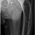
A. Normal wear
B. Hemophilia
C. An infectious process
D. A histiocytic response
2 Which of the following connective tissue disorders has soft tissue calcifications as a prominent manifestation of the disease on radiographs?
A. Rheumatoid arthritis
B. Polymyositis
C. Systemic lupus erythematosus
D. Calcium pyrophosphate dihydrate (CPPD) arthropathy
3a A patient with a history of an inflammatory arthropathy presents for imaging. The provided radiographs demonstrate which of the following findings?
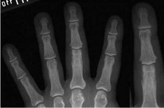
A. Acroosteolysis
B. Sausage digit
C. Gouty tophi
D. Periarticular osteopenia
E. Central “gull wing” erosions
3b Based on the images provided, what is the most likely diagnosis?
A. Rheumatoid arthritis
B. Tophaceous gout
C. Scleroderma
D. Erosive osteoarthritis
E. Psoriatic arthritis
4 A 15-year-old male presents with the images of the feet below. In this disease, what location most commonly undergoes ankylosis?

A. Tarsal bones
B. Carpal bones
C. Hips
D. Lumbar spine
5 What typical findings of calcium pyrophosphate dihydrate (CPPD) arthropathy are noted in the image below?
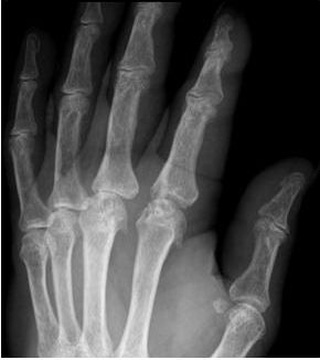
A. Joint space narrowing and marginal erosions
B. Bone demineralization and central erosions
C. Large subchondral cysts and hooked osteophytes
D. Enthesophytes and eburnation
6 What disease process is depicted in the images below?
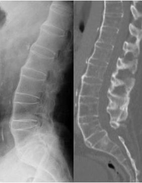
A. Ankylosing spondylitis
B. Diffuse idiopathic skeletal hyperostosis
C. Psoriatic arthritis
D. Reactive arthritis
7 The imaged metabolic disease manifests as an arthropathy characterized by excessive intra-articular and periarticular deposition of what substance?
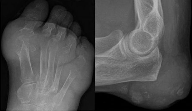
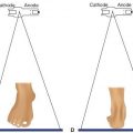
A. Calcium pyrophosphate dihydrate crystals
B. Hemosiderin
C. Monosodium urate crystals
D. B2 microglobulin
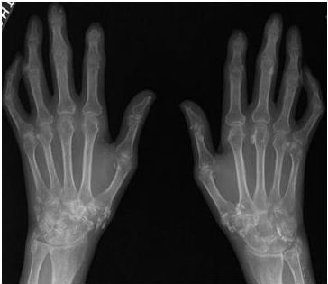
8 Bilateral hand and wrist radiographs are pictured below. What additional imaging finding, outside of the hands and wrists, may also be seen in this same disease process?
A. Esophagram demonstrating achalasia
B. High-resolution chest CT revealing changes of lymphangioleiomyomatosis
C. AP pelvis showing bilaterally symmetric sacroiliitis
D. Small bowel follow-through showing dilated small bowel with closely spaced, thin folds
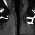
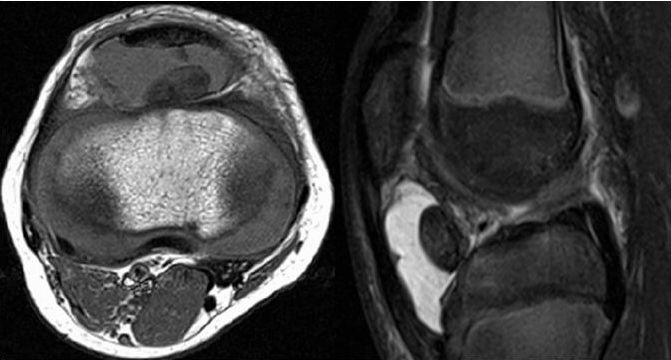
9 Axial T1-weighted and sagittal T2-weighted fat-saturated MR images demonstrate a mass-like focus within the anterior joint space of the knee. Of the choices below, which is the most likely diagnosis?
A. Amyloidosis
B. Pigmented villonodular synovitis
C. Osteochondromatosis
D. Rheumatoid arthritis
10 An 81-year-old male presents with left hip pain. A mass is noted in the expected location of the iliopsoas bursa on the images below. If aspiration of this mass is performed, what would the aspirate most likely resemble?
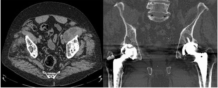
A. Gelatinous material
B. Purulent material
C. Bloody material
D. Serous material
11 A hand radiograph of a patient with rheumatoid arthritis is provided. Which of the following correctly describes the early erosions seen in this disease process?
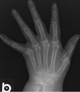
A. Periarticular at the interphalangeal joints and first carpometacarpal joint of the hand
B. Juxta-articular at the first metatarsophalangeal joint in the foot
C. Marginal at the ulnar styloid process and carpal bones of the wrist
D. Central at the metacarpophalangeal joints of the hands
12 The CT image of the pelvis below is most characteristic of which process?
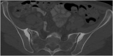
A. Osteitis condensans ilii
B. Ankylosing spondylitis
C. Sacroiliac joint septic arthritis
D. Reactive arthritis
E. Enteropathic sacroiliitis
13 What is the characteristic distribution of osteoarthritis?
A. Metacarpophalangeal joints and distal radioulnar joints
B. Proximal interphalangeal and pisotriquetral joints
C. Intercarpal and index finger metacarpophalangeal joints
D. Distal interphalangeal and thumb carpometacarpal joints
14 A 35-year-old female presents with the following radiographs of the right hand and wrist. What is the most likely diagnosis?
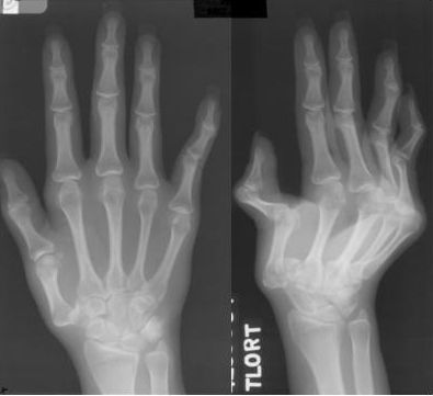
A. Progressive systemic sclerosis
B. Systemic lupus erythematosus
C. Psoriatic arthritis
D. Calcium pyrophosphate dihydrate (CPPD) arthropathy
15 A 55-year-old patient presents with the following radiograph of the lumbar spine. What is the most likely diagnosis?
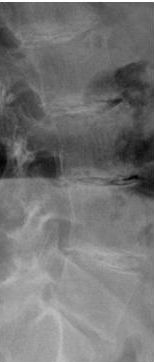
A. Degenerative disc disease
B. Hemochromatosis
C. Ochronosis
D. Spondylitis
16 The image of the hand below demonstrates acroosteolysis. Which disease process has imaging findings including acroosteolysis?
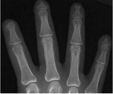
A. Mixed connective tissue disease
B. Calcium pyrophosphate dihydrate (CPPD) arthropathy
C. Rheumatoid arthritis
D. Ankylosing spondylitis
17 A radiograph of the index, middle, and ring fingers is presented. Asymmetric, nonerosive soft tissue swelling around the index finger proximal interphalangeal joint in a patient with underlying osteophytosis is characteristic of what deformity?
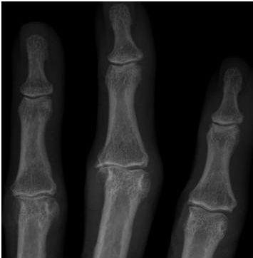
A. Boutonnière deformity
B. Mallet finger
C. Sausage finger
D. Bouchard node
18 The numerous joint and synovial findings seen in the lateral radiograph of the knee are most consistent with which pathology?
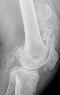
A. Synovial osteochondromatosis
B. Osteoarthritis
C. Myositis ossificans
D. Pigmented villonodular synovitis
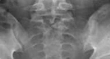
19 A 35-year-old female presents with the following radiograph of the sacroiliac joints. Her radiograph of the lumbar spine, not presented, demonstrates thin, vertical syndesmophytes. What is the most likely diagnosis?
A. Psoriatic arthritis
B. Crohn disease
C. Reactive arthritis
D. Septic arthritis
20a Which of the following is a component of the classic triad of symptoms in reactive arthritis?
A. Cholecystitis
B. Colitis
C. Skin calcifications
D. Urethritis
20b Which of the following genitourinary infections is classically associated with reactive arthritis?
A. Chlamydia trachomatis
B. Treponema pallidum
C. Neisseria gonorrhoeae
D. Trichomonas vaginalis
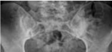
21 In seronegative spondyloarthropathies, where do erosions and sclerosis typically present in the sacroiliac joints?
A. Sacral side of the inferior sacroiliac joint
B. Iliac side of the inferior sacroiliac joint
C. Sacral side of the superior sacroiliac joint
D. Iliac side of the superior sacroiliac joint
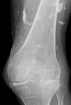
22 The skin calcifications in the image below are typical of which disease process?
A. Calcium pyrophosphate dihydrate (CPPD) arthropathy
B. Dermatomyositis
C. Psoriatic arthritis
D. Systemic lupus erythematosus
23 Calcium pyrophosphate dihydrate (CPPD) deposition arthropathy of the hands and wrists presents with bilaterally symmetric subchondral cysts, beak-like osteophytes, and chondrocalcinosis in a proximal distribution. These findings are similar to which other arthropathy?
A. Reactive arthritis
B. Rheumatoid arthritis
C. Hemochromatosis
D. Gout
24 The posterior lumbar facets are synovial-lined joints. Which part of the lumbar facet joint first shows changes of degeneration?
A. Inferior articulating process
B. Spinous process
C. Ligamentum flavum
D. Superior articulating process
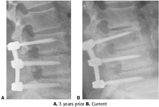
25 Radiographs of the lumbar spine, 3 years prior and current, are presented for evaluation. Which of the following complications has occurred in the interval?
A. Hardware fracture
B. Degenerative disc disease
C. Compression fracture
D. Spondylolysis
26 Which of the following may occur in connective tissue disease?
A. Tumoral calcinosis
B. Calcific tendonitis
C. Hydroxyapatite deposition
D. Calcinosis circumscripta
27 Which of the following arthropathies manifests as cartilage loss in the weight-bearing portions of a joint, bone proliferation, and subchondral cyst formation?
A. Psoriatic arthritis
B. Osteoarthritis
C. Reactive arthritis
D. Rheumatoid arthritis
28 A thoracic spine radiograph is presented below, what is the most likely diagnosis?
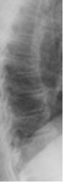
A. Rheumatoid arthritis
B. Psoriatic arthritis
C. Diffuse idiopathic skeletal hyperostosis
D. Ankylosing spondylitis
29 The abnormally increased subchondral bone mineral density in this patient is most likely the result of which process?
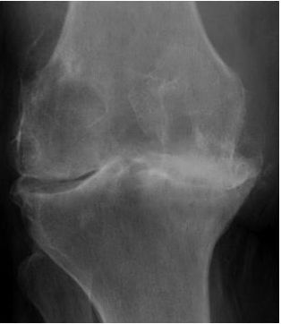
A. Fluorosis
B. Osteoarthritis
C. Paget disease
D. Renal osteodystrophy
30a What arthropathy is noted involving the thumb carpometacarpal joint in the radiographs shown below?
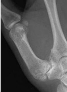
A. Reactive arthritis
B. Rheumatoid arthritis
C. Osteoarthritis
D. Ankylosing spondylitis
30b What are the bony excrescences that extend from the trapezium in this patient?
A. Osteophytes
B. Enthesophytes
C. Syndesmophytes
D. Erosions
31 What inflammatory arthropathy finding is most pronounced in the images below?
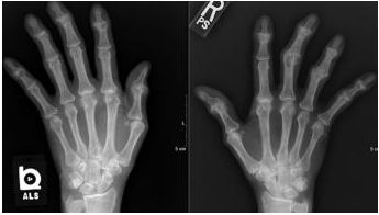
A. Central erosions
B. Periarticular osteopenia
C. Periostitis
D. Subchondral cysts
32 Periostitis is seen in which of the following arthropathies?
A. Psoriatic arthritis
B. Rheumatoid arthritis
C. Gout
D. Osteoarthritis
33 What is the direction of joint space narrowing in this patient?
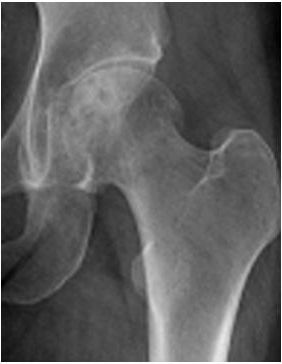
A. Superior
B. Posterior
C. Axial
D. Inferior
34 A 20-year-old male presents with the cervical spine radiograph below. The findings noted in the cervical spine most commonly occur at what level?
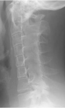
A. C1–C2
B. C2–C3
C. C4–C5
D. C5–C6
35 In this image of a medial compartment prosthesis, which of the following complications is present?
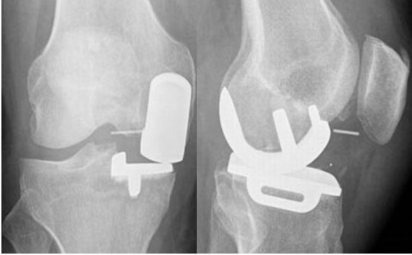
A. Hardware loosening
B. Infection
C. Spacer displacement
D. Fracture
36 Progressive great toe osteoarthritis consisting of osteophyte formation, joint space narrowing, and subchondral sclerosis can lead to what condition?
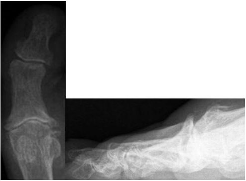
A. Hallux rigidus
B. Hallux valgus
C. Turf toe
D. Morton syndrome
37 What is the diagnosis based on the provided AP radiograph of the bilateral feet?
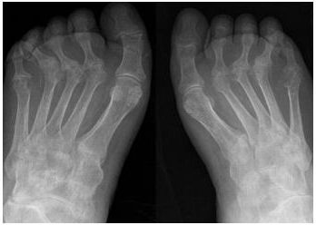
A. Ankylosing spondylitis
B. Psoriatic arthritis
C. Osteoarthritis
D. Rheumatoid arthritis
E. Gout
38 What is the earliest radiographic manifestation of the arthropathy depicted in the hands and wrists below?
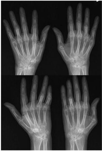
A. Soft tissue swelling and juxta-articular osteoporosis
B. Erosions of the ulnar styloid and metacarpal heads
C. Radiocarpal and interphalangeal joint space narrowing
D. Metacarpophalangeal joint subluxation
39 What is the most likely etiology of the findings at the L3–L4 level on the CT image below?
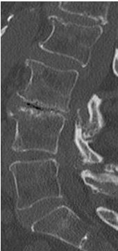
A. Ankylosing spondylitis
B. Degenerative disc disease
C. Discitis–osteomyelitis
D. Diffuse idiopathic skeletal hyperostosis
40 Which of the following radiographic findings is the most sensitive for the detection of arthroplasty loosening?
A. Any bone cement lucency
B. Change in the position of the hardware
C. Stress shielding
D. Polyethylene component wear
ANSWERS AND EXPLANATIONS
1 Answer D. Particle disease is also known as aggressive granulomatosis. It is a histiocytic response of the bone to small polyethylene particles shed from the lining of the hardware. It causes localized osteolysis and may demonstrate a superiorly malpositioned femoral head component. The provided image demonstrates a large lucency around the femoral component of the arthroplasty, consistent with osteolysis. Treatment commonly requires revision. The collection around the hip can be distinguished clinically from infection by lack of night pain and fever, but hip aspiration may be required to confirm the diagnosis.
Reference: Garcia GM. Musculoskeletal Radiology. New York, NY: Thieme; 2010:66.
2 Answer B.
Stay updated, free articles. Join our Telegram channel

Full access? Get Clinical Tree


