A. Soft tissue abscess
B. Joint effusion
C. Chronic osteomyelitis
D. Myositis
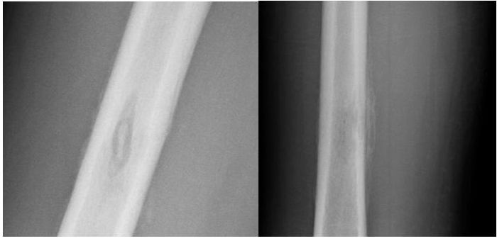
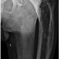
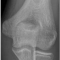
2a A 26-year-old male is transferred from an outside institution after a radiograph at that institution was interpreted as a possible malignancy of the right femur. Based on the radiographs below, what is the most likely diagnosis?
A. Osteosarcoma
B. Ewing sarcoma
C. Metastatic disease
D. Osteomyelitis
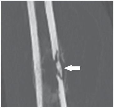
2b A CT examination of the previous finding was performed. What is indicated by the arrow in the coronal CT image below performed in bone window?
A. Sequestrum
B. Involucrum
C. Periosteal reaction
D. Sinus tract
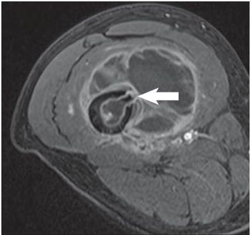
2c An MRI of the above finding was performed. What is indicated by the arrow on the axial T1 fat-saturated postcontrast image below?
A. Sequestrum
B. Involucrum
C. Cloaca
D. Periosteum
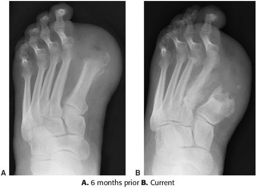
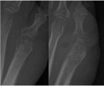
3 Pictured below are two radiographs of a diabetic right foot, one current and one 6 months prior immediately after amputation of a portion of the great toe. In diabetic patients, what is the most common site of osteomyelitis of the foot?
A. Navicular and medial cuneiform
B. Third and fourth metatarsal shafts
C. Cuboid and lateral cuneiform
D. First and fifth metatarsal heads
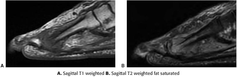
4 A 7-year-old female presents to the emergency department with ankle swelling. MR images of the distal tibial and fibular metaphyses are presented. What is the most likely diagnosis?
A. Necrotizing fasciitis
B. Subperiosteal abscess
C. Pyomyositis
D. Gas gangrene
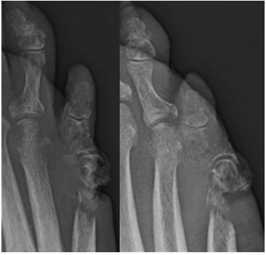
5 Images of the fifth metatarsal–phalangeal joint are presented in a 49-year-old patient. Which of the following is the earliest radiographic finding of septic arthritis?
A. Osteomyelitis
B. Joint effusion
C. Periarticular osteoporosis
D. Erosions
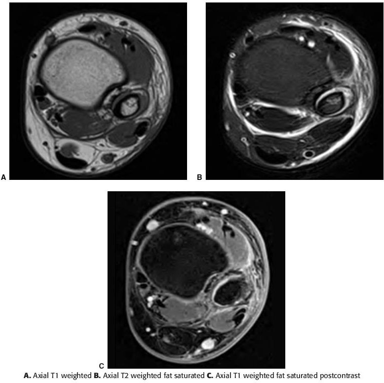
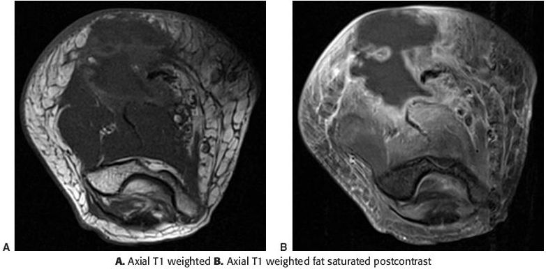
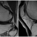
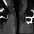
6 A 25-year-old male presents to the emergency department complaining of left hip pain. Based on the MR images below, what is the diagnosis?
A. Cellulitis
B. Pyomyositis
C. Osteomyelitis
D. Necrotizing fasciitis
7 Presented below are two MR images of osteomyelitis of the second metatarsal in a diabetic patient. Which T1-weighted signal pattern is most reliable in diagnosing osteomyelitis of the foot?
A. Low geographic, medullary T1 signal
B. Low hazy, reticulated T1 signal
C. Low subcortical T1 signal
D. Low subchondral T1 signal
8 A 24-year-old female presents with swelling of the elbow and forearm. What is the characteristic appearance of the fibrous capsule of an abscess on MRI?
A. Thin, nonenhancing
B. Thin, enhancing
C. Thick, nonenhancing
D. Thick, enhancing
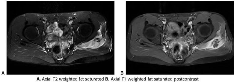
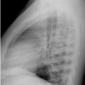
9 A 65-year-old male presents to the emergency room with a fever, malaise, and bilateral posterior thigh pain. A CT of the bilateral lower extremities was performed. Which of the following microorganisms is most often implicated in the condition depicted on the CT image?
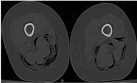
A. Salmonella
B. Staphylococcus
C. Streptococcus
D. Polymicrobial
10a A 59-year-old diabetic male presents with the following images of the right foot. What is the earliest osseous radiographic sign of osteomyelitis?
A. Permeative osseous destruction
B. Endosteal scalloping
C. Indistinctness of the cortex
D. Periosteal reaction
10b The ordering physician is concerned about gangrene of the foot. Which of the following MRI findings is most critical in diagnosing gangrene of the foot?
A. Signal voids resulting from gas created by gas-forming organisms
B. Skin thickening and cellulitis
C. Areas of absent contrast enhancement
Related posts:
Stay updated, free articles. Join our Telegram channel

Full access? Get Clinical Tree


