A. Freiberg infraction
B. Insufficiency fracture
C. Köhler disease
D. Lisfranc injury
2 What is the most common carpal coalition?
A. Scapholunate
B. Scaphotrapeziotrapezoidal
C. Lunotriquetral
D. Capitate–hamate
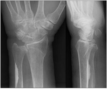
3 The radiograph demonstrates what wrist abnormality?
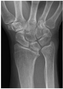
A. Scapholunate advanced collapse (SLAC wrist)
B. Kienböck disease
C. Perilunate dislocation
D. Madelung deformity
4 Plexiform neurofibromas are most commonly seen in which of the following syndromes?
A. Neurofibromatosis type 1
B. Neurofibromatosis type 2
C. Sturge-Weber
D. Tuberous sclerosis
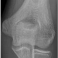
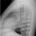
5a A 30-year-old male presents with the following radiograph. The abnormality most commonly results in which one of the following clinical symptoms?
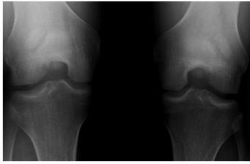
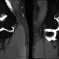
A. Anterior knee pain
B. Lateral knee pain
C. Inability to fully extend the knee
D. Inability to fully flex the knee
5b What is the most common MRI finding in this anomaly?
A. Quadriceps tendinosis
B. Patellar tendinosis
C. Hyperintense T2 signal within the bipartite fragment
D. Nonenhancement of the bipartite fragment suggesting devitalized bone
6 Which one of the following clinical manifestations is associated with the syndrome depicted by the CT and radiograph below?
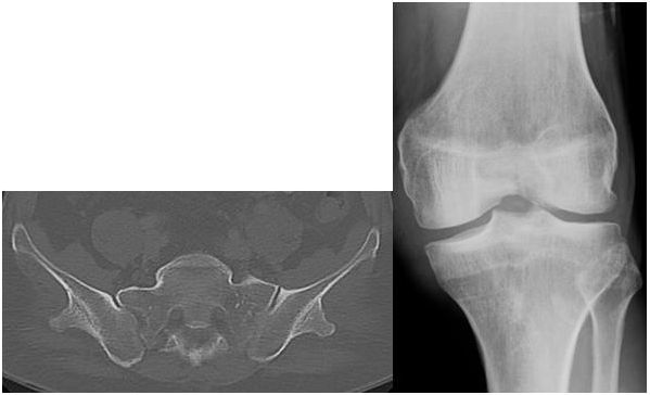
A. Ataxia
B. Nail changes
C. Hypertension
D. Arrhythmias
7 Which of the following would be included in the differential diagnosis for the findings noted in the image below?
A. Congenital insensitivity to pain
B. Hypoparathyroidism
C. Hyperthyroidism
D. Osteoarthrosis
8 Bridging ossification between osseous structures in fibrodysplasia ossificans progressiva typically occurs where initially?
A. Pelvis
B. Sternocleidomastoid
C. Shoulder girdle
D. Upper arms
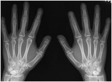
9 A patient presents with enlargement of the second and third digits of the right hand. On radiographs of the right hand, there is osseous overgrowth of the second and third digit, long and broad phalanges, splayed distal tufts, bowing deformity, and enlargement of the soft tissues. What is the most likely diagnosis?
A. Dermatomyositis
B. Scleroderma
C. Macrodystrophia lipomatosa
D. Leprosy
10 Which of the findings below is more commonly seen in primary hypertrophic osteoarthropathy as compared to secondary hypertrophic osteoarthropathy?
A. Phalangeal tuft hypertrophy
B. Phalangeal tuft acroosteolysis
C. Phalangeal tuft sparring
D. Phalangeal tuft sclerosis
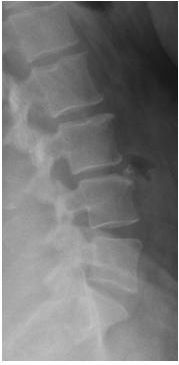
11 A 21-year-old male presents with back pain. Which of the following is correctly associated with the diagnosis depicted by the image below?
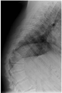
A. Disc space widening
B. Anterior wedging of at least four consecutive vertebral bodies
C. Decreased AP diameter of the vertebral bodies
D. Schmorl nodes
12 A 69-year-old female presents with neck pain and limited mobility. What is the most likely diagnosis?
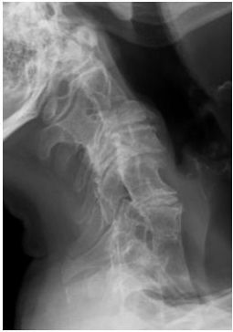
A. Juvenile idiopathic arthritis
B. Sprengel deformity
C. Klippel-Feil syndrome
D. Prior surgical fusion
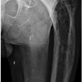
13 A patient presents with chronic elbow pain. How is congenital dislocation of the radial head distinguished from a prior traumatic radial head dislocation?
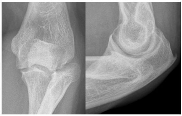
A. The trochlea is dysplastic with congenital dislocation of the radial head.
B. There is a fracture line in the radial head with congenital dislocation of the radial head.
C. There is a persistent effusion associated with congenital dislocation of the radial head.
D. The radial head is overgrown and dysplastic in congenital dislocation of the radial head.
14 A 10-year-old female presents with the image below. This patient most likely has which type of this disease?
A. Type I
B. Type II
C. Type III
D. Type IV
15 A 78-year-old female presents with wrist pain. What is the most common location for the finding in the ulna?
A. Upper extremity
B. Lower extremity
C. Spine
D. Skull
16 Which of the following statements about the movement of an os odontoideum on flexion and extension views is correct?
A. It moves with the atlas on flexion but not extension.
B. It moves with the axis on flexion but not extension.
C. It moves with the atlas on flexion and extension.
D. It moves with the axis on flexion and extension.
17 What is the primary etiology leading to the entity depicted in this pelvis radiograph?
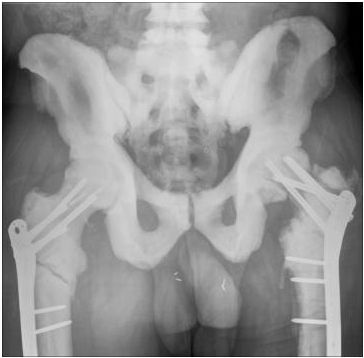
A. Abnormal osteoclast function
B. Abnormal osteoblast function
C. Abnormal calcium distribution
D. Abnormal phosphate distribution
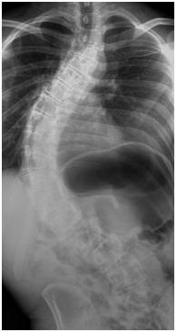
18 A 38-year-old female presents with back pain. The finding at L4 most likely represents which of the below options?
A. Syndesmophyte
B. Compression fracture
C. Gibbus deformity
D. Limbus vertebra
19 A 20-year-old male presents with scoliosis, as noted in the image below. What is the most common cause of scoliosis?
A. Idiopathic
B. Trauma
C. Leg length discrepancy
D. Congenital
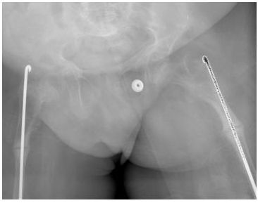
20 A 17-year-old female presents with left hip pain resulting from developmental dysplasia of the hip. Which of the following radiographic findings is consistent with developmental dysplasia of the hip?
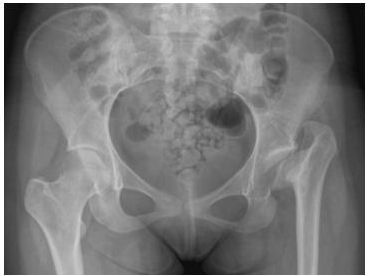
A. Downturning of the lateral acetabular roof
B. Coxa magna
C. Distal displacement of the trochanters
D. Anteversion of the acetabulum
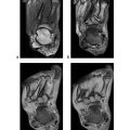
21 An 18-month-old presents with scoliosis. Based on the three consecutive sagittal T2-weighted fat-saturation images, what is the most likely cause of scoliosis in this patient?
Related posts:
Stay updated, free articles. Join our Telegram channel

Full access? Get Clinical Tree


