A. Hyperthyroidism
B. Hyperparathyroidism
C. Hypothyroidism
D. Hypoparathyroidism
2b What is the earliest radiographic finding of hyperparathyroidism?
A. Subperiosteal resorption of the medial cortex of the tibia
B. Subchondral resorption of the distal clavicle
C. Subligamentous resorption of the coracoclavicular ligament at the clavicular attachment
D. Subperiosteal resorption of the radial surface of the second and third digit middle phalanx
2c What percentage of cases of primary hyperparathyroidism results from a parathyroid adenoma?
A. 75% to 85%
B. 55% to 65%
C. 35% to 45%
D. 15% to 25%
3 In acromegaly, a heel pad thickness greater than how many centimeters is characteristic for a male?
A. 1.8 cm
B. 2.3 cm
C. 2.8 cm
D. 3.3 cm
4a A 75-year-old female with osteoporosis presents with pain in the right hip. What is most likely responsible for the finding below?
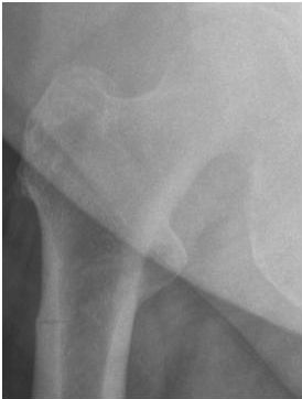
A. Looser zone
B. Bisphosphonate fracture
C. Traumatic fracture
D. Stress fracture
4b What is the next most appropriate test?
A. Radiograph of the left hip/femur
B. Radiograph of the thoracic/lumbar spine
C. MRI of the right hip/femur
D. MRI of the pelvis/sacroiliac joints
4c What is the best imaging test to diagnose osteoporosis?
A. Radiographic skeletal survey
B. MRI of the sacrum
C. DEXA scan
D. MRI of the lumbar spine
5 A 58-year-old male with renal osteodystrophy presents with the following radiograph. What crystal is responsible for the calcifications?
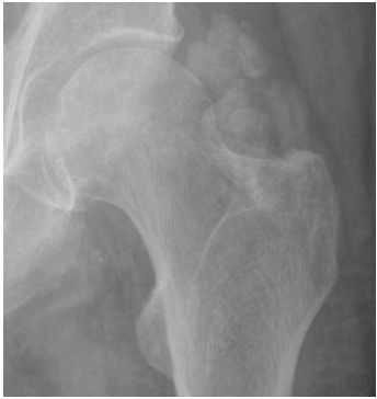
A. Calcium pyrophosphate
B. Uric acid
C. Calcium hydroxyapatite
D. Sodium urate
6 What is the most common location for periosteal new bone formation in thyroid acropachy?
A. Tibia
B. Humerus
C. Distal phalanges
D. Metacarpals
7 In myelofibrosis, which of the following is a characteristic bone marrow MRI appearance?
A. T1-weighted bone marrow signal is lower than muscle or intervertebral disc signal.
B. T2-weighted and STIR bone marrow signal is higher than normal.
C. Bone marrow enhances on postcontrast images.
D. Marrow signal drops out on opposed-phase imaging compared to in-phase imaging.
8 Which radiographic finding is seen in the setting of thalassemia?
A. Thickened cortex that causes a “hair-on-end” appearance of the skull.
B. Diffuse osteosclerosis of the bones of the axial skeleton.
C. Widened medullary space of the tubular bones leads to absence of tubulation.
D. Thinned horizontal trabeculae of the spine lead to a striated vertebral body appearance.
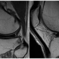
9a Based on the image below, what is the most likely cause of the visualized findings?
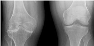
A. Chondrocalcinosis resulting from hyperparathyroidism
B. Chronic osteomyelitis associated with S. aureus infection
C. Disuse osteopenia resulting from a recent ipsilateral ankle fracture
D. Hemarthrosis in the joint resulting in synovitis
9b After the knee, what is the next most common joint involved in hemophilia?
A. Shoulder
B. Elbow
C. Hip
D. Ankle
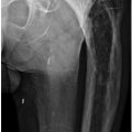
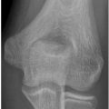
10 The finding below is classically associated with which disease?
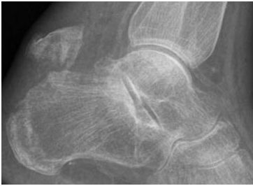
A. Hyperparathyroidism
B. Renal osteodystrophy
C. Diabetes mellitus
D. Osteomalacia
11a Which of the below statements is the most accurate internal standard for evaluating the MRI appearance of red marrow?
A. The T1-weighted signal of red marrow is greater than skeletal muscle or intervertebral discs.
B. The T1-weighted signal of red marrow is less than skeletal muscle or intervertebral discs.
C. The T2-weighted signal of red marrow is greater than skeletal muscle or intervertebral discs.
D. The T2-weighted signal of red marrow is less than skeletal muscle or intervertebral discs.
11b Which portion of the bone converts red marrow to yellow marrow last?
A. Epiphysis
B. Apophysis
C. Metaphysis
D. Diaphysis
11c Which of the following is the most characteristic imaging appearance of mass-like extramedullary hematopoiesis?
A. Homogeneous nonfatty soft tissue masses in the appendicular skeleton
B. Homogeneous nonfatty soft tissue masses in the axial skeleton
C. Heterogeneously fatty soft tissue masses in the appendicular skeleton
D. Heterogeneously fatty soft tissue masses in the axial skeleton
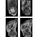
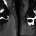
12a A 34-year-old male presents with back and pelvic pain. What is the most likely cause of the bone marrow signal pattern?
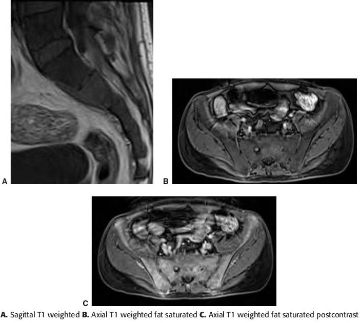
A. Marrow reconversion
B. Sickle cell anemia
C. Aplastic anemia
D. Malignancy
12b A 21-year-old male presents with hip and pelvic pain. What is the most likely cause of the bone marrow signal pattern?
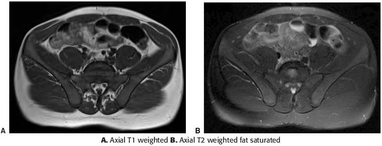
A. Marrow reconversion
B. Sickle cell anemia
C. Aplastic anemia
D. Malignancy
12c A 20-year-old college athlete presents with right shoulder pain after a recent increase in training before the beginning of the season. What is the most likely cause of the bone marrow signal pattern?
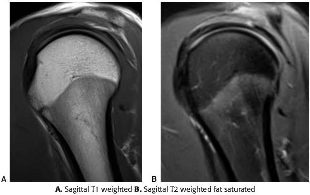
A. Marrow reconversion
B. Sickle cell anemia
C. Aplastic anemia
D. Malignancy
13
Related posts:
Stay updated, free articles. Join our Telegram channel

Full access? Get Clinical Tree


