Artifacts
Mark Doyle
OVERVIEW
Successful identification of artifacts is crucial to allow correct interpretation of image features. Because image formation is dependent on numerous acquisition and patient features, there are consequently multiple manifestations of image artifacts. Major sources include the following:
Motion
Blood flow
Susceptibility
Fat and water resonance differences
Incorrect imaging parameters
MOTION
To encode each k-space line requires application of two separate gradients:
Frequency encoding (X)
Phase encoding (Y)
The imaging gradients relate the frequency of spins at each location to position within the image. When the signal is sampled, the main feature of the spins that determines their position in the image is how the signal phase progresses throughout the two-dimensional (2D) k-space domain. K-space has the property that motion of an object is efficiently represented as a change of phase in the k-space signal. For instance, the k-space representation of a ball bouncing around the magnetic resonance imaging (MRI) scanner visually looks the same for each position of the ball (see Fig. 36-1). For each frame, the phase of the k-space signal is different. To appreciate the origin of this, consider a spin moving along a magnetic field gradient: it experiences a progressively increasing precessional frequency as it moves into a progressively increasing magnetic field. In this way, a moving spin will accumulate phase at a different rate compared to a static spin. Therefore, following a gradient echo operation, a moving spin will accumulate a net phase that is due in part to its position and its motion. However, because phase information is used in k-space to encode position, it follows that moving spins will appear at irregular positions within the image and that these positions do not merely reflect the new position that they moved to. Spin motion results in artifacts because the Fourier transform does not have enough information to correctly assign the spins to one position within the image. The type of motion that takes place determines the form of the motion artifact:
Cyclic motion—cyclic repeating artifact
Irregular motion—random, dispersed signal artifact
Slow motion—confined, localized blurring of object
Breathing Artifact
When performing rapid imaging, the motion of the diaphragm can be appreciated because the diaphragm moves approximately 5 cm during regular breathing. The cyclic respiratory motion is typically regular and this property usually causes respiratory artifacts to reinforce as regions of displaced signal within the image (see Fig. 36-2). Not surprisingly, moving features such as internal organs and the chest wall generate signals that propagate throughout the image. Their regular motion tends to make these moving features appear as if they were in two positions on average and produce a displaced “ghost image.” In images acquired under free breathing conditions, a strong respiratory motion artifact is typically seen as a “ghost,” that is, a displaced image chest wall and lung cavity. Other, possibly fainter, features are typically present also. Scout images are widely regarded as “throw away images,” and are usually acquired under free breathing and possibly noncardiac triggered conditions. Naturally, unless some form of respiratory compensation is used, these images
will be of lower quality, have less distinct boundaries, and be contaminated by artifact. Commonly, breath-hold imaging is employed to reduce or eliminate respiratory artifacts. Table 36-1 summarizes some features that differentiate images acquired under breath-hold and free breathing conditions.
will be of lower quality, have less distinct boundaries, and be contaminated by artifact. Commonly, breath-hold imaging is employed to reduce or eliminate respiratory artifacts. Table 36-1 summarizes some features that differentiate images acquired under breath-hold and free breathing conditions.
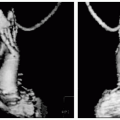
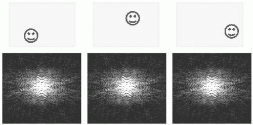 FIGURE 36-1 The top panel shows a ball bouncing around inside the magnetic resonance imaging (MRI) scanner, three snapshots are shown. The corresponding k-space maps are shown below the images. Visually, there is nothing to distinguish one k-space map from another. This illustrates that motion of a real object (i.e., the ball) is represented in the phase of the k-space signal. To visualize phase, as opposed to magnitude, requires additional image processing. |
Respiratory Gating
An alternative to breath-holding is to employ respiratory gating. Respiratory gating requires some means of monitoring the diaphragm position within the respiratory cycle and adjusting the scan acquisition accordingly. Strategies of adjusting data acquisition include the following:
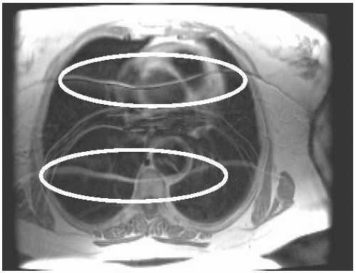 FIGURE 36-2 This image was acquired under free breathing conditions and displays common breathing artifacts. Coherent ghost images of the chest wall are seen repeating through the image (circled). |
Not acquiring data during periods of high motion
Tracking the mobile feature and applying real-time adjustment of the imaging slice
Acquiring regions of k-space that have higher or lower sensitivity to motion in a manner that is dependent on the details of motion
Pulsatile Blood Flow
A noncardiac triggered scan tends to represent pulsatile blood vessels as regularly repeating patterns. This form of artifact is especially problematic for time of flight (TOF) 2D angiographic imaging, because the frequency of the pulsatile blood flow is highly organized relative
to the acquisition and imposes a definite pattern in the phase information over successive k-space lines. The repeating ghost vessels tend to be very prominent (see Fig. 36-3).
to the acquisition and imposes a definite pattern in the phase information over successive k-space lines. The repeating ghost vessels tend to be very prominent (see Fig. 36-3).
TABLE 36-1 Features of Images Acquired Under Breath-Hold and Free Breathing Conditions | ||||||||
|---|---|---|---|---|---|---|---|---|
|
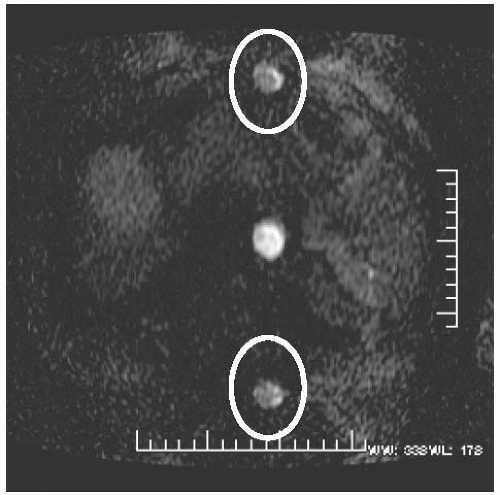 FIGURE 36-3 Pulsatile flow artifacts are shown (circled). This image was taken as part of a time of flight scan. The scan time was rapid relative to the pulsatile blood cycle, and hence the spacing of the ghost images of the vessel are seen relatively widely spaced. |
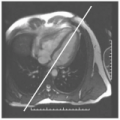
Direction of Motion Artifact
Motion artifact exhibits a preferential direction within the image and occurs in the phase encoding direction. Consider that the measurement gradient is applied for only a short time duration relative to most motion features whereas the phase encoding gradient is applied at multiple times throughout the motion cycle. Therefore, the phase encoding process is more highly disrupted by motion. For this reason, motion artifacts predominantly occur as displacements along the direction of the phase encoding gradient (see Fig. 36-4).
Rapid Scout Imaging
There are three features that are problematic when using nontriggered, non-breath-hold scout images:
Respiratory artifact
Cardiac motion artifact
Incorrect position for planning future breath-hold images
Related posts:
Stay updated, free articles. Join our Telegram channel

Full access? Get Clinical Tree


