George M. Bridgeforth and George Holmes
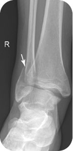
A 46-year-old woman falls on her right ankle while ice-skating.
CLINICAL POINTS
- Bimalleolar fractures are fractures of the distal fibula and tibia.
- Trimalleolar fractures are fractures of the distal fibula, tibia, and the posterior lip of the distal tibia.
- About one-third of all ankle fractures are bimalleolar and trimalleolar fractures.
Clinical Presentation
Bimalleolar and trimalleolar fractures account for more than 30% of all ankle fractures. Patients complain of marked pain, swelling, and tenderness associated with an inability to bear weight or ambulate. The mechanism of injury is direct impact caused by inversion or eversion injuries. Inversion injuries occur more often. The non–weight-bearing distal fibula is thinner than the distal tibia. In addition, the lateral collateral ligaments (the anterior and posterior talofibular ligaments and the posterior talofibular ligament) are thinner than the stronger broad-based medial collateral (deltoid) ligament. Eversion injuries are less common, but they are more likely to result in unstable fractures.
Pain, swelling, and tenderness along either or both malleoli may be secondary to an acute fracture or a severe ankle sprain. However, the presence of a distal tibial fracture or a disrupted deltoid ligament (marked swelling 5 mm or greater between the talus and the distal tibia) should prompt the examiner to search for a fracture of the distal fibula or torn lateral collateral ligaments. The physician should
- Perform the anterior and posterior drawer tests to check for ankle instability
- Check for varus and valgus instability
- Check for tenderness along the posterior edge of the tibia posterior tibia (posterior tibial fracture)
- Palpate the fifth metatarsal for an associated metatarsal fracture, the navicular for focal tenderness for possible fracture, and the talus (check for talar pain with ankle dorsiflexion) for an associated talar fracture
- Inspect for heel pain (calcaneal fracture)
- Determine whether there is neurovascular impairment (cold, cyanotic limb with diminished or absent pulses, impaired sensation). It is essential to document the neurovascular examination carefully.
PATIENT ASSESSMENT 
- Marked pain, swelling, and tenderness
- Limited range of motion
- Difficulty weight-bearing with an inability to take four steps.
Radiographic Evaluation
Anteroposterior (AP), lateral, and oblique views should be ordered. Additional tests to order include the following:
- Mortise view: the ankle is rotated internally 10 to 15 degrees. A mortise view eliminates the overlap of the distal fibula and tibia seen on routine AP projections and allows for better visualization of the talar dome and the distal tibia (plafond). It may be used as part of acute trauma series for high-impact injuries. Uses include detection of pilon fractures of the distal tibia or fractures of the talar dome as well as osteochondritis dissecans of the tibiotalar joint. This latter condition appears as a minute semioval (saucer-shaped) bite mark along the joint line. Osteochondritis dissecans should be suspected in any patient who complains of joint locking or joint mice.
- Stress views: usually obtained by specialists. These views place an anatomical load on the ankle mortise. An increase in the space between the talus and the surrounding fibula or tibia 5 mm or more is suspicious for a complete disruption of the lateral or medial collateral ligament. In addition, a malunion in the anatomical relationship may indicate an old fracture (it is also necessary to look for bony sclerosis). Many specialists prefer weight-bearing views, which may be very useful; however, patients with severe foot and ankle injuries may have problems weight-bearing.
NOT TO BE MISSED 
- Open ankle fractures
- Ankle contusion
- Associated fifth metatarsal fracture (Jones vs. styloid)
- Associated talar fracture
- Septic joint
- Gout
- Psoriatic arthritis
Related posts:


Stay updated, free articles. Join our Telegram channel

Full access? Get Clinical Tree


