George M. Bridgeforth and John Cherf
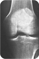
A 26-year-old man fell and bruised his right knee. He now complains of pain, swelling, and tenderness. (From Bucholz RW, Heckman JD. Rockwood & Green’s Fractures in Adults. 5th ed. philadelphia, PA: Lippincott Williams & Wilkins; 2001.)

CLINICAL POINTS
- The reported incidence of bipartite patella may be as high as 6%.
- The condition occurs more often in men.
- Most cases are bilateral.
Clinical Presentation
Bipartite patella is a congenital condition in which the patella is actually two separate bones, not one, as is usually the case. The cause is incomplete fusion of the ossification centers of the patella. Generally, individuals do not know that they have the disorder. Most cases are asymptomatic; incidental findings on radiographs confirm the presence of the condition. At least 1% of people are affected, and the reported incidence may
be as high as 6%. The condition is nine times more common in men than in women and is usually unilateral.
A bipartite patella rarely causes pain and functional limitations that require treatment. Direct trauma to the patella or repetitive injury may trigger symptoms. Common manifestations include pain directly over the patella, swelling at the synchondrosis, and pain-limited range of motion of the knee.
Radiographic Evaluation
It is necessary to order anteroposterior (AP), lateral, and oblique views. Optional radiographs include the following:
- Full trauma series (for serious traumatic injuries)
- Oblique views (two): used on a full trauma series to assess for condylar fractures
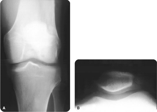
- Sunrise or Merchant view: used to check for subtle patella dislocations and vertical patella fractures (Fig. 19.1). A Merchant view is more sensitive for detecting patella subluxations.
- Intercondylar view: used to check for tibial spine and tibial plateau fractures
PATIENT ASSESSMENT 
- Usually asymptomatic
- Radiographic finding
- Knee pain following trauma or overuse
Usually, a bipartite patella appears as a large rounded bone fragment, which is generally located in the upper outer quadrant. It is best seen on the AP view (Fig. 19.2). Occasionally, it may be misdiagnosed as a fracture. This confusion may be important for medicolegal reasons (see “Treatment”). However, unlike an acute fracture, a bipartite patella usually has smooth but parallel margins. One researcher has noted that when a bipartite patella appears as a separate bony island, it may have sclerotic margins. Another has pointed out that the pieces of a bipartite patella do not fit together exactly, like a puzzle, but that the separate bone fragments from a fracture do appear to fit together like the pieces of a puzzle.

FIGURE 19.1 Radiographic appearance of a bipartite patella on anteroposterior (A) and Merchant (B) views of the knee. (Courtesy of Dr. Rebecca Loredo.) (From Bucholz RW, Heckman JD. Rockwood & Green’s Fractures in Adults. 5th ed. Philadelphia, PA: Lippincott Williams & Wilkins; 2001.)
Stay updated, free articles. Join our Telegram channel

Full access? Get Clinical Tree


