George M. Bridgeforth and George Holmes
A 47-year-old man fell off a ladder and landed on his feet. He complains of marked pain, swelling, and tenderness in his left foot.
CLINICAL POINTS
- Most calcaneal fractures are unilateral.
- 10% rule:
- 10% of the injuries are bilateral.
- 10% may be associated with burst fractures of the spine.
- 10% may be associated with a compartment syndrome.
- 10% of the injuries are bilateral.
- These fractures are sometimes associated with compression fractures of the spinal vertebrae.
Clinical Presentation
Calcaneal fractures account for approximately 2% of all fractures seen in adults. These fractures most often occur in young men. The calcaneus has three subtalar articular facets—the anterior, middle, and posterior facets—that support the talus. The fourth articular facet articulates with the cuboid of the midfoot. Approximately 33% of all calcaneal fractures are extra-articular and include all fractures that do not involve the posterior facet. Approximately 67% of calcaneal fractures are intra-articular fractures, which are calcaneal fractures that involve all four articular facets.
Most calcaneal fractures are caused by direct trauma—direct impact resulting from falls from heights or high-impact motor vehicle collisions (Fig. 29.1). When evaluating calcaneal fractures, it is important to remember the 10% rule:
- 10% of calcaneal fractures are bilateral (Fig. 29.2).
- 10% (approximately) are associated with burst fractures of the thoracolumbar region. It is essential to examine both feet and the spinal column carefully for pain and tenderness, bruising, and swelling.
- 10% (approximately) of affected individuals may have a compartment syndrome (marked swelling of the extremity, resulting in neurovascular compromise). When conducting a neurovascular examination, it is important to always check for swelling, coldness, and pallor of the ankle and the foot. The posterior tibial arterial pulses may be detected along the posterior medial malleolus. The dorsal pedis arterial pulses lie just medially to the extensor hallucis tendon at the midfoot. It is necessary to ensure that capillary refill is 2 seconds or less. Patients at risk for developing compartment syndrome should be referred and kept under observation.
Patients may complain of pain, bruising, and swelling as well as an inability to bear weight on the injured foot. They frequently have an impaired heel strike during weight bearing. The examiner should check the contralateral heel to exclude the presence of bilateral injury. Also, it is necessary to inspect and palpate other areas at high risk for fracture, such as the bony part of the ankle, as well as the metatarsals. Patients with marked comminuted calcaneal fractures may exhibit a heel deformity, but this finding is absent with nondisplaced fractures.
It is also important to document the integrity of the Achilles tendon. Patients with an intact Achilles tendon have a normal Thompson’s test. A normal test is characterized by plantar flexion (downward toward the floor) of the foot when the calf is squeezed by the examiner.
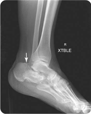
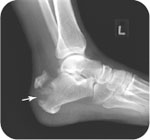
FIGURE 29.1 Lateral radiograph of the right calcaneus in a 47-year-old man involved in a motor vehicle collision, demonstrating an acute fracture from an axial load on the calcaneus.
As previously stated, calcaneal fractures may be accompanied by compression fractures of the spinal vertebrae. The axial load from falls may cause a burst fracture in the lower thoracic/upper lumbar spine. Although uncommon, lower burst fractures involving L5 have been reported in the literature. Therefore, patients who are seen in acute settings following major falls should have an assumed spinal cord injury until proven otherwise. Patients with burst fractures commonly exhibit marked focal tenderness over the fracture site. Neurological findings with acute burst fractures of the lower thoracic/upper lumbar region are variable. The findings may range from no functional neurological deficits to acute paraplegia at T10 (the umbilicus) with neurological damage (lower motor neuron injury), impairing bowel and bladder function. See Chapter 5 for more information about burst fractures.
Radiographic Evaluation
Anteroposterior (AP), lateral, oblique, and calcaneal views should be ordered. For acute occult fractures, computed tomography (CT) scans are necessary. For ruptures of the plantar fascia, magnetic resonance imaging (MRI) scans should be obtained (Fig. 29.3).
Boehler’s angle helps identify a calcaneal fracture. One line is drawn from the posterior tubercle to the apex of the calcaneus. Another line is drawn from the anterior tubercle to the apex of the calcaneus. The angle formed in front of the two intersecting lines is the Boehler’s angle. An angle of less than 25 degrees should prompt suspicion for an underlying calcaneal fracture.
PATIENT ASSESSMENT 
- Pain and tenderness of the heel
- Inability to bear weight (on the heel)
- Deformity of the heel or plantar arch
Related posts:


Stay updated, free articles. Join our Telegram channel

Full access? Get Clinical Tree


