Fig. 1
Axial T2 sequence without fat suppression for staging of rectal cancer. The mesorectal fat surrounds the rectum and is enclosed within the mesorectal fascia (yellow arrows). In the left panel, the tumor was staged as an early T3 tumor with minimal invasion into the perirectal fat. The distance from the mesorectal fascia is more than 1 cm (red arrow). In the middle panel, a more extensive example of a T3 tumor is shown with a tumor that approaches within 2 mm of the mesorectal fascia (large white arrow). In the right panel, a sagittal view is shown. A mesorectal lymph node is visible (thin white arrow). The estimated distance of the tumor from the anal verge is 4.5 cm
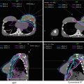
Rectal tumors are well visualized on PET, so PET/CT is also becoming a standard part of staging and planning to help delineate the extent of gross disease (Fig. 2). However, areas of low uptake on PET should not supercede physical exam findings or abnormalities seen on CT.


Fig. 2
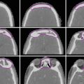
A patient with T4N0 rectal adenocarcinoma (invasion into the cervix). GTV: These panels illustrate the utility of PET in target volume delineation. In the upper panel, the GTV (red) is seen on representative axial, sagittal, and coronal images, respectively, on both the treatment planning CT and PET. The lower panel shows additional axial slices
3 Simulation and Daily Localization
Most patients that are to be treated with standard 3D conformal radiotherapy are simulated in the prone position with the use of a belly board for anterior displacement of the bowel. However, if IMRT is planned, then we recommend that the patient be simulated in the supine position in a body mold or other immobilization device to ensure more accurate setup reproducibility. A radiopaque marker should be placed on the anus.
CT simulation with ≤3 mm thickness with IV contrast should be performed to delineate the pelvic blood vessels and gross tumor volume. The use of oral contrast may be helpful to identify the small bowel, which is an important organ at risk. If PET/CT is available, a PET/CT fusion should be obtained to aid in target volume delineation. For patients that underwent preoperative MRI, MR fusion could also aide with treatment planning.
Bladder filling/emptying may be considered, especially if IMRT is used. A full bladder may keep bowel from migrating into the pelvis. An empty bladder may be more reproducible.
We recommend daily orthogonal kilovoltage images and weekly cone beam CT scans (to assess soft tissue) to verify alignment during treatment. Cone beam CTs may be done more frequently if there is significant variation in bladder and/or rectal filling.
4 Target Volume Delineation and Treatment Planning
Conventional 3D conformal radiotherapy for rectal cancer involves a PA field and two opposed lateral fields (Figs. 3 and 4).

Fig. 3
Standard fields used for a T3N1 rectal cancer receiving preoperative chemoradiation, PA (left panel) and left lateral (right panel). The CTV-SR is shown in red. The patient was simulated in prone in a belly board allowing the small bowel (shown in purple) to fall anteriorly away from the CTV. The bladder is shown in yellow

Fig. 4
Standard fields used for a T3N2a rectal cancer receiving postoperative chemoradiation following an APR. PA (left panel) and left lateral (right panel). The CTV-SR is shown in red. The field includes the perineal scar. The patient was simulated in prone in a belly board allowing the small bowel (shown in purple) to fall anteriorly away from the CTV. Note that more small bowel is in the pelvis in the postoperative setting
Traditional borders for the PA field are superior, L5/S1 interspace; inferior, the inferior edge of the obturator foramen or 3 cm below the GTV, whichever is more distal; and lateral, 1.5–2 cm beyond the pelvic brim.
Borders for the lateral fields include superior, same as PA field; inferior, same as PA field; anterior, posterior margin of the pubic symphysis (bony landmark for internal iliac nodes) for T3 disease or at least 1 cm anterior to the anterior edge of the pubic symphysis (bony landmark for external iliac nodes) for T4 disease; and posterior, 1–1.5 cm posterior to the posterior edge of the bony sacrum.
With the use of CT-based planning, the borders described above can be modified to ensure adequate coverage of the planning target volumes (PTV). Target volumes including gross tumor volume (GTV), clinical target volumes (CTV), and the PTV should be delineated on every applicable slice of the planning CT.
The primary gross tumor volume (GTV-P) is defined as all gross disease on physical examination and imaging.
The nodal GTV (GTV-N) includes all visible perirectal, mesorectal, and involved iliac lymph nodes. Include any lymph node in bout as GTV-N in the absence of a biopsy.
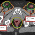
The high-risk CTV (CTV-HR) should include the GTV with a minimum of 1.5–2-cm superior and inferior margin as well as the entire rectum, mesorectum, and presacral space. Additional details are given in Table 1.
Table 1
Suggested target volumes for gross and microscopic disease in the preoperative setting
Target volumes
Definition and description
GTV (gross tumor volume)
Primary (GTV-P): all gross disease on physical examination and imaging
Regional nodes (GTV-N): all visible perirectal and involved iliac nodes; include any lymph node in doubt as GTV in the absence of a biopsy
CTV-high risk (CTV-HR)
CTV-HR should cover the GTV-P and GTV-N with 1.5–2-cm margin expansion superiorly and inferiorly, but excluding the uninvolved bone, muscle, or air. This volume should include the entire rectum, mesorectum, and presacral space axially at these levels. A 1–2-cm margin around gross tumor invasion into adjacent organs should be added. Coverage of the entire presacral space and mesorectum should be strongly considered. Any visible mesorectal nodes on CT and PET should also be included
A 1.8-cm-wide volume between the external and internal iliac vessels is needed to cover the obturator nodes (Taylor et al. 2005)
CTV-standard risk (CTV-SR)
Should cover the entire mesorectum and right and left internal iliac lymph nodes for T3 tumors. The right and left external iliac lymph nodes for T4 tumors with anterior organ involvement should also be included
A 1–2-cm margin in adjacent organs with gross tumor invasion should be added for T4 lesions
Superiorly, the entire rectum and mesorectum should be included (usually up to L5/S1) and at least 2-cm margin superior to gross disease, whichever is most cephalad
Inferiorly, the CTV should extend to the pelvic floor or at least 2 cm below the gross disease, whichever is most caudad
A 1.8-cm-wide volume between the external and internal iliac vessels is needed to cover the obturator nodes (Taylor et al. 2005)
Planning target volume (PTV)
Each CTV should be expanded by 0.5–1 cm, depending on the physician’s comfort level with setup accuracy, frequency of imaging, and the use of IGRT
The standard-risk CTV (CTV-SR) should cover the entire mesorectum and bilateral internal iliac lymph nodes for T3 tumors. The CTV-SR should also include the bilateral external iliac nodes for patients with T4 tumors with anterior organ involvement. Additional details are found in Table 1.
Target volume delineation in the postoperative setting is similar to the preoperative setting. However, in the case of an abdominoperineal resection, the entire surgical bed extending inferiorly to the level of the perineal scar needs to be included. Additional details are given in Table 2.
Table 2
Suggested target volumes in the postoperative setting
Target volumes
Definition and description
CTV (positive margin or gross disease)
Should include the area of known microscopically involved margin or macroscopic residual disease plus a 1–2-cm margin, but exclude the uninvolved bone, muscle, or air
CTV-high risk (CTV-HR)
Should include the entire remaining rectum (if applicable), mesorectal bed, and presacral space axially at these levels but exclude uninvolved bone, muscle, or air. Coverage of the entire presacral space and mesorectum should be considered
CTV-standard risk (CTV-SR)
Should cover the entire mesorectum and right and left internal iliac lymph nodes for T3 tumors. The right and left external iliac lymph nodes for T4 tumors with anterior organ involvement should also be included
Superiorly, the entire remaining rectum and mesorectum should be included (usually up to L5/S1) and at least 1-cm margin superior to the anastomosis, whichever is most cephalad
Inferiorly, the CTV should extend to the pelvic floor or at least 1 cm below the anastomosis or rectal stump, whichever is most caudad. If patient is status post an abdominoperineal resection, the surgical bed extending down to the perineal scar should be included. The scar should be outlined with a radiopaque marker.
To cover the external iliac nodes (for T4 lesions), an additional 1-cm margin anterolaterally around the vessels is needed. Any adjacent small nodes should be included (Myerson et al. 2009; Taylor et al. 2005)
Stay updated, free articles. Join our Telegram channel

Full access? Get Clinical Tree

 Get Clinical Tree app for offline access

