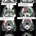
Fig. 1
Functional pelvic anatomy determining vulvar cancer resectability: T2-weighted pelvic MRI with water-based endoluminal vaginal gel (a) and corresponding CT (b) imaging highlighting important pelvic structures integral in determining the functional resectability of vulvar cancers
For most vulvar cancers involving the labia and vestibule, the first echelon lymphatic drainage is to the superficial medial inguinal and deep femoral lymph nodes. The inguinal-femoral lymph nodes are an anatomically defined compartment laterally bound by the medial border of the iliopsoas, medially by the lateral border of the adductor longus, posteriorly by the iliopsoas muscle, and pectineus and anteriorly by the edge of the sartorius muscle and rectus femoris (Kim et al. 2012) (see Fig. 2).

Fig. 2
Inguinal-femoral lymph node compartment: anatomical compartment for the first echelon inguinal-femoral lymph nodes showing the muscles defining the compartment
Vulvar lymphatic channels do not cross the labiocrural fold and generally do not cross the midline for well-lateralized lesions >1 cm from midline.
Pelvic lymph nodes mainly represent second echelon lymphatics only affected when inguinal-femoral nodes are involved, the exception being small lymphatics of midline structures (clitoris or bulb of the vestibule) that drain directly to pelvic lymphatics via the internal pudendal and iliac lymphatics. As seen in GOG 37, 28 % of patients with inguinal node positivity had involvement of pelvic lymph nodes.
A prior MRI study with injected iron oxide particles showed that 95 % of the common iliac, internal iliac, medial and anterior external iliac, and obturator lymph nodes are located with 7 mm of the pelvic vasculature (Taylor et al. 2005). Conversely, for inguinal-femoral lymph nodes and the lateral external iliac region, variability of body habitus and prior lymph node dissection limits the applicability of the vessel surrogate plus 7 mm definition, where involved lymph nodes range from 0.9 to 3.5 cm from the vessel, and thus the anatomical compartment rather than fixed circumferential should be used in defining inguinal nodes (Kim et al. 2012).
2 Diagnostic Workup Relevant for Target Volume Delineation
Initial physical examination should include a comprehensive gynecologic exam with specific attention paid to noting the local extent of the disease and inguinal nodal involvement. In GOG 37, 23.8 % of patients with clinically enlarged lymph nodes were found to have no evidence of disease on pathologic assessment, while 23.9 % of patients with a normal exam were found to have occult metastatic disease. Thus physical examination is neither sensitive nor specific in identifying the extent of nodal involvement. CT or PET-CT is therefore essential in workup and treatment planning.
Extrapolating from other pelvic malignancies, most prominently anal cancer, where the role of PET-CT is well established, we recommend incorporating PET-CT-based planning for all vulvar patients to increase the sensitivity of identifying nodal disease that would otherwise be undetected by the combination of physical exam and CT imaging (Cotter et al. 2006).
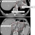
The use of gadolinium-enhanced pelvic MRI with and without water-based endoluminal vaginal gel is important in locally advanced disease to evaluate the relationship between tumor and functionally integral normal tissues such as the musculature of the anal sphincter and vagina that preclude surgical resection (see Fig. 3).

Fig. 3
MR imaging of the locally advanced vulvar cancers. (a, b) Posterior vulvar lesion (highlighted by the arrow) extending to the anus and puborectalis muscle. (c, d) Anterior vulvar lesion with extensive urethral and preurethral involvement (highlighted by the arrow)
When disease involves the vagina, a gold fiducial marker is placed during in-office speculum examination to identify the vaginal extent of disease aiding in GTV definition and daily setup.
3 Simulation and Daily Localization
CT or PET-CT simulation in the supine position with the lower extremities abducted (“frog-legged”) and immobilized in a vacuum-evacuated device to assure reproducibility in setup. By abducting the lower extremities to the maximal extent allowable by the scanner, the dose to the medial thigh and groin folds is minimized.
In order to assist in delineation of the tumor, radiopaque wire is used to identify areas of gross disease or the postoperative tumor bed (see Fig. 4).

Fig. 4
Radiopaque wire for simulation. PET-CT based simulation with gross disease outlined by a radiopaque wire placed at the time of simulation (highlighted by the arrows), corresponding to the PET avid disease seen on simulation imaging
CT or PET-CT simulation using 3–5 mm thickness with IV and oral contrast. Oral contrast assists in the small bowel and rectum visualization, while the IV contrast aids in visualization of the vasculature structures for nodal contouring. We typically recommend a simulation scan from at least the L2 through the mid thigh. The isocenter is typically placed in the center of the pelvis if treating lymph nodes. In comparison, it is typically placed in the vulva at the level of pubic symphysis if targeting only the vulva.
If the vagina is involved (and thus to be included in CTV1 as described below), we suggest performing the simulation with a full and empty bladder to create an ITV (internal target volume) for the vaginal region. It is recommended that all vulvar patients be treated with a full bladder and empty rectum irrespective of vaginal involvement. On the day prior to simulation, patients are instructed to take a fleet enema and are re-simulated if rectal distention is >3.0–3.5 cm since an empty rectum represents the most conservative posterior location for rectal filling effects on interfraction motion.
We avoid the use of a vaginal obturator or marker as these devices distort anatomy and create non-reproducible anatomical deformation of the pliable vaginal mucosa. Additionally, the devices displace the vagina posteriorly, creating a situation prone to geographic miss.
A customized bolus of 0.5–1 cm thickness is usually created for the vulvar region at the time of treatment planning and is used for daily treatments. Separate plans are generated with and without bolus at the time of initial IMRT planning, such that should the patient develop a brisk skin reaction, treatment can be continued without bolus and no attendant treatment interruption. Additionally a 1–2 cm thick bolus structure is extended in air for IMRT plan optimization only, which serves to extend the isodose line beyond the skin for flash to account for edema and swelling (see Fig. 5). The difference in monitor units with and without bolus should be minimal.

Fig. 5
Customized bolus for IMRT optimization to generate flash. CT treatment planning images with and without a customized bolus structure generated for IMRT optimization to extend isodose lines beyond the vulva generating a flash of dose beyond the skin (highlighted by arrow)
Table 1
Suggested IMRT dose fractionation schemes used for vulvar cancer
Clinical situation
PTV1 (Gy)
PTV2 (Gy)
Dose per fraction
Preoperative GOG 205 (Moore et al. 2012)
45–50.4
55.8–59.4
1.8 Gy daily
Definitive
45–50.4
59.4–70.2
1.8 Gy daily
Adjuvant
45–50.4
50.4–59.4
1.8 Gy daily
Patients are typically set up with a combination of daily KV and CBCT imaging. Daily kV imaging is used to reduce interfraction motion based upon the bony anatomy or to fiducial markers placed at the vaginal extent of disease if present. For boost CBCT daily would be preferred for daily localization and matching with the soft tissue.
4 Target Volume Delineation and Treatment Planning
The primary GTV includes all gross disease on physical examination and imaging. For pelvic and inguinal, GTV includes all lymph nodes ≥1 cm in the short axis, those with a necrotic center and/or PET avidity (see Table 1 and 2).
Table 2
Suggested target volumes at the high-risk subclinical region
Target volumes
Definition and description
GTV
Primary: All gross disease on physical examination and imaging (see above regarding the importance of PET-CT and MRI)Related posts:

Stay updated, free articles. Join our Telegram channel

Full access? Get Clinical Tree

 Get Clinical Tree app for offline access