Fig. 1
(a) T1-weighted sagittal sequence. (b) T1-weighted axial sequence
Anteriorly, the tumor can extend into the nasal fossa (87 %) (Fig. 7) and result in destruction of the pterygoid plates (27 %). Less commonly, the tumor can invade the ethmoid and maxillary sinus or infiltrate the orbital apex.
Laterally, the tumor can extend into the parapharyngeal space (68 %), which is an important part of T staging for a tumor (Figs. 7 and 8b). Extension into this space can occasionally lead to invasion of cranial nerves IX to XII in advanced disease.
Superiorly, nasopharyngeal carcinoma can directly invade the base of the skull, the sphenoid sinus, and the clivus (41 %). The foramen lacerum is a vulnerable spot through which the tumor may enter the cavernous sinus (16 %) and the middle cranial fossa to invade cranial nerves III to VI. The foramen ovale also allows access for the tumor to invade the middle cranial fossa, in addition to the petrous portion of the temporal bone (19 %), and the cavernous sinus.
The posterior extension of NPC is less common and can include invasion of prevertebral muscles (19 %) and inferior invasion of the oropharynx (21 %).
Ipsilateral lymphadenopathy is appreciable in 85–90 % of NPC cases, and bilateral disease is present in approximately 50 % of cases.
Retropharyngeal lymph nodes are commonly involved in nasopharyngeal cancer (Fig. 3b).
Lypmh node chains routinely involved include levels II–V. Level IA is rarely invovled with the disease. Figure 2 shows the frequency of detection of lymph nodes at various levels in NPC based on an MR study (Ng et al. 2007).

2 Diagnostic Workup Relevant for Target Volume Delineation
Fiber-optic examination provides the best means to assess the mucosal spread of disease. Typically, lesions are exophytic in nature; however, 10 % may be submucosal and not visible on endoscopic examination.
Cranial nerve deficits can occur in 10 % of patients with NPC, and their detection will result in alterations to target volumes (most commonly CN VI, V, XII, IX, and X).
For CN IX and X: Cover jugular foramen
For CN V: Cover pterygopalatine fossa, cavernous sinus, foramen rotundum, and foramen ovale
For CN VI: Cover cavernous sinus
MRI provides superior assessment of skull base involvement and tumor invasion into soft tissue structures compared to CT scan (Abdel Khalek Abdel Razek & King 2012), in particular.
T2-weighted fast spin echo: Depict involvement of parapharyngeal space, paranasal sinus, and retropharyngeal lymph node involvement (Fig. 3b).
T1 non-contrast: Determine involvement of clivus (sagittal cuts) and skull base involvement (Fig. 3c). Tumor invasion of marrow is typically hypointense relative to normal marrow.

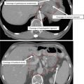
Fig. 3
MR imaging of the nasopharynx: (a) T1-weighted post-contrast sequence demonstrates a right-sided nasopharyngeal primary invading the prevertebral musculature. Note that the fascial plane demarcating the parapharyngeal space is intact (arrow). (b) T2-weighted fat-saturated sequence demonstrating a left-sided retropharyngeal lymph node (arrow). Often on CT, these can be difficult to distinguish clearly from the primary. (c) Sagittal T1-weighted sequence demonstrates diffuse infiltration of the marrow of the clivus (arrow)
T1 post-contrast: Determine perineural invasion and intracranial involvement (Fig. 8).
Fusion of the skull base portion of the MRI can aid in the delineation of the GTV. CT still provides superior assessment of cortical bone involvement.
MRI is better able to differentiate retropharyngeal lymph nodes from the primary tumor compared to CT.
Any evidence of enlarged retropharyngeal lymph nodes should be considered gross disease. For other lymph node beds, the presence of central necrosis, extracapsular spread, or a short-axis diameter of 10 mm suggests possible involvement with the disease. PET/CT may also help clarify whether borderline nodes are involved with the disease. As a general rule, given that NPC has a high likelihood of nodal spread, any suspicious nodes should be considered as gross disease.
3 Simulation and Daily Localization
Set up the patient in supine position with the head extended. The immobilization device should include at least the head and neck. If possible, shoulders should also be immobilized to ensure accurate patient setup on a daily basis especially when an extended-field IMRT plan is used. A bite block can be placed during simulation and throughout radiation to push the tongue away from the high-dose nasopharynx region.
CT simulation using 3 mm thickness with IV contrast should be performed to help guide the GTV target, particularly for the lymph nodes. We typically recommend a simulation scan from the top of the head including the brain to the carina. 5 mm thickness can be reconstructed below the clavicle to the level of the carina. The isocenter is typically placed just above the arytenoids.
Image registration and fusion applications with MRI and PET scans should be used to help in the delineation of target volumes, especially for regions of interest encompassing the GTV, skull base, brainstem, and optic chiasm. The GTV and CTV and normal tissues should be outlined on all CT slices in which the structures exist.
Daily image-guided setup with KV images can allow for reduced margins near critical structures (brainstem), and we often utilize this in our own clinical practice.
4 Target Volume Delineation and Treatment Planning
Suggested target volumes at the GTV and high-risk CTV are detailed in Tables 1 and 2 and are based on guidelines from RTOG Nasopharyngeal Cancer Trials (Lee et al. 2009). Alternative volume delineation strategies and dose fractionation schemes are thoroughly discussed in a review article by Wang et. al (Wang et al. 2012). Selected fractionation schemes are briefly reviewed in Table 3.
Table 1
Suggested target volumes at the gross disease region
Target volumes
Definition and description
GTV70 a (the subscript 70 denotes the radiation dose delivered)
Primary: all gross disease on physical examination and imaging (see above regarding the importance of MRI)
Neck nodes: all nodes ≥ 1 cm or those with necrotic center
CTV70 a
PTV70 a
CTV70 + 3–5 mm, depending on the comfort level of daily patient positioning. Around critical structures like the brainstem, a 1 mm margin is acceptable (Fig. 10)
Table 2
Suggested target volumes at the high-risk subclinical region
Target volumes
Definition and description
CTV59.4 a
CTV59.4 (Figs. 4, 5, 6, 7, 8, 9, 10, and 11) should encompass CTV70 with a 5 mm margin and regions at risk for microscopic disease which include:Get Clinical Tree app for offline access


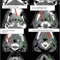
Fig. 4
Target delineation GTV70 (red) and CTV59.4 (green) in a patient with T1N1 nasopharyngeal carcinoma with retropharyngeal and level II nodes in a cranial-to-caudal direction. Notice the inclusion/exclusion of nodal level 1B of the N + (green) versus N0 (blue) neck. Please note that these are representative slices and not all slices are included