Chapter 63 Embryology of the heart is covered in Chapter 62. In this chapter, some embryologic events that are fundamental to understanding the segmental approach to heart disease are reiterated. The heart develops from two simple epithelial tubes that fuse to form a single tube (Fig. 63-1) with the following components: Figure 63-1 Bulboventricular looping of the primitive heart tube may occur to the right (D-looping) or to the left (L-looping). Sinus venosus consists of right and left horns. Each horn receives blood from three important veins: the umbilical vein, the common cardinal vein, and the vitelline vein. Paired primitive atria will later fuse to form a common atrium. The atrioventricular (AV) sulcus divides the common atrium and the primitive ventricle. The primitive ventricle becomes the left ventricle (LV). The interventricular sulcus divides the primitive ventricle and the bulbus cordis. The bulbus cordis may be divided as follows: the proximal third gives rise to the body of the right ventricle (RV). The distal-most section is the truncus arteriosus, which develops into the aortic root and part of the pulmonary artery (PA). The remaining mid portion is the conus cordis, which connects the primitive RV to the truncus arteriosus. The conus cordis partitions to form the outflow tracts of the RV and LV. Although the two ends of the heart tube remain relatively fixed, rapid growth of the middle section results in the development of a large S-shaped curve called the bulboventricular loop (see Fig. 63-1). As the heart tube grows and becomes longer, it usually bends to the right, termed D-looping by Van Praagh. D-looping is responsible for the proximal bulbus cordis (RV) lying anterior and to the right of the primitive ventricle (LV). If the heart tube loops to the left, termed L-looping, the RV will lie anterior and to the left of the LV. In the heart tube stage, the primitive LV and the proximal bulbus cordis (primitive RV) are separated from the truncus arteriosus (which gives rise to both great arteries) by the conus or infundibulum. The conus consists of the subpulmonary and subaortic conus cushions. Normally, expansile growth of the subpulmonary conus occurs, causing it to protrude anteriorly on the left, carrying the pulmonary valve anteriorly, superiorly, and to the left of the aortic valve. Resorption of the subaortic conus occurs. Hence the aortic valve lies posterior, inferior, and right-sided, in direct fibrous contiguity with the mitral valve (Fig. 63-2). The anterior pulmonary artery rises above the anterior ventricle (RV) and leads to the posterior sixth arterial arch, which forms the branch pulmonary arteries. The posterior aorta originates above the posterior LV and leads to the anterior fourth arterial arch (which forms the aortic arch). Figure 63-2 Normal and abnormal conal development. 1. The development of the suprahepatic portion of the inferior vena cava (IVC) is closely linked to the growth of the liver, and thus the anatomic right atrium (RA) and the liver almost invariably develop on the same side of the body. This concept of visceroatrial situs is fundamental to the segmental approach. 2. Ventricular looping is independent of the visceroatrial situs. This phenomenon gives rise to the concept of concordance (RA-RV and left atrium [LA]-LV) and discordance (RA-LV and LA-RV). 3. Ventricular looping and the great arterial relationship are independent entities. The direction of bulboventricular looping and the development of the conotruncus are responsible for the ultimate relationship of the great arteries to each other and to the underlying ventricles and AV valves. One can think of the heart as a three-level house (Fig. 63-3). The first level is the visceroatrial situs, the middle level is the ventricular loop, and the third level is the conotruncus. To describe it simply, the three levels are the atria, ventricles, and great arteries. The heart has two staircases: the AV ventricular junction and the ventriculoarterial junction. The levels represent the major cardiac segments. The staircases represent the connecting segments. Figure 63-3 The “house” model of the heart. The segmental approach to heart disease comprises the following steps: 1. What is the anatomic type of each of the three major cardiac segments: the atria, the ventricles, and the great arteries? 2. How is each segment connected to the adjacent segment? 3. What are the associated anomalies involving the valves, atrial and ventricular septum, the great vessels, and the systemic and pulmonary veins? 4. How do the segmental combinations and connections, with or without the associated malformations, function? The defining features of the morphologic RA (systemic venous atrium) and LA (pulmonary venous atrium) are based on their venous connections, as well as their appendage and pectinate muscle morphology. Using venoatrial connections for atrial identification is based on the fact that the sinus venosus, which carries the systemic venous return, is an integral part of the morphologic RA. Hence the morphologic RA receives the IVC and the superior vena cava (SVC) and the orifice of the coronary sinus. However, the SVC and coronary sinus have a high incidence of variation, which can be a source of diagnostic confusion. These variations include left SVC to an unroofed coronary sinus and bilateral SVC with the left SVC draining to an unroofed coronary sinus. In these cases, the SVC would appear to drain into the LA. In rare instances, even the IVC may drain into the coronary sinus, which may be unroofed, or the coronary sinus septum may be absent. In spite of this rare exception, the most reliable means of identifying the morphologic RA by cross-sectional imaging is by recognizing its connection to the IVC (Fig. 63-4). Even in the setting of an interrupted IVC, a suprahepatic segment of the IVC is present entering the RA, allowing accurate identification. Figure 63-4 The inferior vena cava (IVC) as a marker of the morphologic right atrium (RA). 1. Muscular connection between the free wall and the interventricular septum (moderator band) (Fig. 63-5, A) Figure 63-5 Right ventricular identification.
Cardiovascular Anatomy and Segmental Approach to Imaging of Congenital Heart Disease
Embryologic Basis for the Segmental Approach to Heart Disease
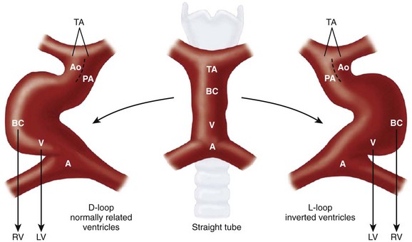
Ao, aorta; BC, bulbus cordis; LV, left ventricle; PA, pulmonary artery; RV, right ventricle. (Modified from Van Praagh R, Weinberg PM, Matsuoka R, et al. Malposition of the heart and the segmental approach to diagnosis. In: Adams FH, Emmanouilides GC, eds. Moss’ heart diseases in infants, children and adolescents. 3rd ed. Baltimore: Williams & Wilkins; 1983.)
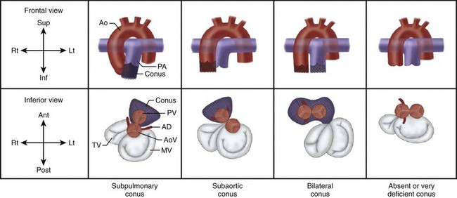
A, Subpulmonary conus seen in normally related great arteries. B, Subaortic conus in typical transposition of the great arteries. C, Bilateral conus, as in double-outlet right ventricle. D, Absent or deficient conus, as in double-outlet left ventricle. On the side of the conus, the semilunar valve sits atop the muscular infundibulum, and no direct fibrous contiguity exists between the semilunar valve and the atrioventricular (AV) valve. On the side of the deficient conus, direct fibrous contiguity usually exists between the AV valve and the semilunar valve. Ant, anterior; Ao, aorta; AoV, aortic valve; Inf, inferior; Lt, left; MV, mitral valve; PA, pulmonary artery; Post, posterior; PV, pulmonary valve; Rt, right; Sup, superior; TV, tricuspid valve. (Modified from Van Praagh R, Weinberg PM, Matsuoka R, et al. Malposition of the heart and the segmental approach to diagnosis. In: Adams FH, Emmanouilides GC, editors: Moss’ heart diseases in infants, children and adolescents. 3rd ed. Baltimore: Williams & Wilkins; 1983.)
Segmental Approach to Diagnosis of Congenital Heart Disease
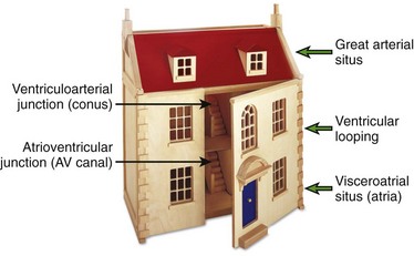
The three levels (major cardiac segments) are the atria, ventricles, and great arteries. The house has two connecting walls with doors (connecting segments): the atrioventricular junction and the ventriculoarterial junction. The house has two entrances: the systemic veins and the pulmonary veins.
Identification of the Major Cardiac Segments
Atrial Identification
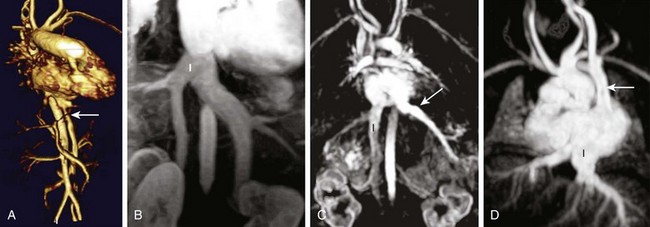
A, The IVC (arrow) and aorta are on the left and the IVC enters the left-sided atrium, which represents the morphologic RA. B, Bilateral IVC that fuse (I) prior to entering the right atrium. C, The right hepatic vein enters the IVC (I), which drains into the RA, while the remaining hepatic veins (arrow) drain separately into the left atrium. D, Left sided-interrupted IVC with azygos continuation. A suprahepatic segment of the IVC (I) drains into the left-sided right atrium in this patient with atrial situs inversus. A left superior vena cava also is present (arrow).
Ventricular Identification
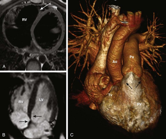
A, Moderator band of the right ventricle (RV) (arrow). B, The atrioventricular (AV) valve of the RV (tricuspid valve) is more apically displaced (arrow) than the AV valve of the left ventricle (LV) (mitral valve) (arrow). C, The conus (arrowsRelated posts:
![]()
Stay updated, free articles. Join our Telegram channel

Full access? Get Clinical Tree


