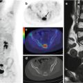
Fig. 33.1
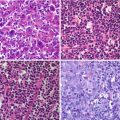
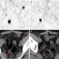
A 19-year-old man with fever, anemia, asthenia. A mesenterial lymph node and several small iliac lymph nodes were identified on ultrasonographic examination and contrast-enhanced CT scan. (a, b) The PET study showed an 18F-FDG-avid mesenterial mass (yellow arrow in a) corresponding to the large lymph node depicted on contrast-enhanced CT. Histopathological analysis showed Castleman’s disease, hyaline-vascular subtype
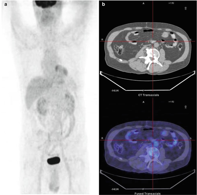
Fig. 33.2
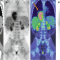
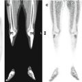
PET study after chemotherapy. Maximum intensity projection (a), CT and PET/CT fusion images (b). Although the lymph nodes are still visible on the morphological exam, its metabolic activity on PET is not significant, suggesting a complete disease response in accordance with the clinical signs
Teaching Point
Castleman’s disease is a rare lymphatic polyclonal disorder characterized by unicentric or multicentric lymph node hyperplasia and nonspecific symptoms and signs, including fever, asthenia, weight loss, an enlarged liver, and abnormally high blood levels of numerous antibodies. Given the high glucose metabolic activity seen in CD, 18F-FDG PET is an appropriate imaging modality to stage or restage the disease and to evaluate the response to treatment.
References
1.
2.
Lin CY, Huang TC (2011) Cervical posterior triangle Castleman’s disease in a child – case report & literature review. Chang Gung Med J 34:435–439PubMed
Stay updated, free articles. Join our Telegram channel

Full access? Get Clinical Tree


