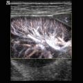
Fig. 12.1
The normal ovarian blood supply. (a) Vascular supply of the ovary by the ascending branch of the uterine artery, the adnexal and tubal branches of the uterine artery and the main ovarian artery. (b) Arterial blood supply of the fallopian tube and ovary and the often tortuous intraovarian arterial branches, which build the vascular arcades for the follicles. (Drawing by Blankvisual, Thun, Switzerland)
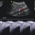
Doppler investigation of the intraovarian arteries shows low flow velocities and low-vascular-resistance-type flow profiles (see Fig. 12.2).

Fig. 12.2
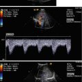
Normal ovarian blood flow. CDI of the normal intraovarian artery in a girl aged 4 months (the ovary is located in an inguinal herniation) shows a normal low-resistance flow profile
After puberty in the inactive ovary (without active follicle), arterial flow shows a high-resistance flow profile. In the active ovary (during ovulation and during the formation of the corpus luteum), the increased blood supply leads to a low-resistance flow profile (RI ~ 0.5) with positive diastolic flow velocities (Golestani et al. 2008).
12.2 Clinical Indications of Paediatric Duplex Sonography of the Ovaries
There are three common indications for the (duplex) sonographic investigation of the ovaries in the paediatric age group:
Abdominal pain in the lower quadrants suggesting ovarian torsion or inflammation or with a clearly lower intensity of pain cystic disease
The evaluation of an ovarian mass seen as an incidental finding in abdominal sonography
The evaluation of suspected inguinal herniation
12.2.1 Ovarian Torsion Versus Inflammation
Adnexal torsion is a common paediatric emergency suggested in many cases of abdominal pain if the most common causes (right-sided pain, appendicitis; left-sided pain, constipation) have been excluded. The diagnosis is often difficult because of the unclear clinical symptoms and findings of the physical examination. A significant proportion (about 15 %) of ovarian torsions occur during childhood in infantile girls.
The greyscale sonographic appearance of ovarian torsion varies and suggests torsion in any case of abnormal shape and size of the ovary.
The ovarian volume can be assessed by the ellipsoid formula (volume = 1/2 length × width × height).
The upper limit of normal for ovarian volume in adults is 15 cm3.
Paediatric ovarian mean volumes are reported as 1 cm3 or less in girls of 6 years and younger, 1.2–2.3 cm3 in girls of 6–10 years and 2–4 cm3 in girls of 11–12 years (Kupesic and Plavsic 2010; Shadinger et al. 2008).
The ovaries of both sides should be evaluated by CDI to compare volume and arterial and venous flow velocity.
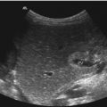
In many cases of ovarian torsion, a cystic structure is seen in the duplex investigation which is often thought to be the cause of the torsion (see Fig. 12.3a).

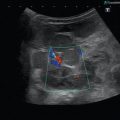
Fig. 12.3
CDI findings in torsion versus inflammation (a) Doppler findings in ovarian torsion: CDI shows no intraovarian arterial and venous Doppler signals in a surgical-proven ovarian torsion in a 12-year-old patient. The volume of the ovary was 20 ml, clearly above the normal range. (b, c) Whirlpool sign in ovarian torsion in a girl aged 4 years. The fallopian tube shows circular hypoechogenic areas around a hyperechogenic centre (b) which can be identified as (venous) vessels by CDI (c). (d) Sagittal investigation of an ovarian torsion in a girl aged 12 years reveals a retrovesical twisted fallopian tube (arrow) and cystic structure in the left ovary which is probably the cause of the torsion. (e) In inflammatory disease CDI reveals a strongly increase vascularity in this 13-year-old girl
Abdominal pain, vomiting, ovarian enlargement and the absence of ovarian venous Doppler flow are the most frequently seen clinical and CDI indicators of ovarian torsion.
The so-called whirlpool sign in the twisted vascular pedicle of the ovary is the most definitive sign of ovarian torsion (Servaes et al. 2007). The whirlpool sign is mostly defined as a circular wrapping of the hypoechoic dilatated venous vessels around the echogenic central axis of the fallopian tube (see Fig. 12.3). Despite its worth, the whirlpool sign is not widely accepted in the European countries, maybe due to the lack of whirlpools here.
Even in the presence of arterial and venous Doppler flow, most authors recommend surgical exploration if the clinical symptoms are as said above and ovarian enlargement is present on duplex investigation (Hurh et al. 2002; Poonai et al. 2013; Servaes et al. 2007; Shadinger et al. 2008).
The finding of a detectable arterial and venous circulation despite ovarian torsion can be explained by the double arterial blood supply of the ovary and possible anatomic variations in the blood supply.
Typical duplex findings in inflammatory disease are an, often slight, enlargement and an increased perfusion of the ovary (see Fig. 12.3).
MRI is, by some centres, used as an additional diagnostic tool in suspected ovarian torsion; in our experience, the loss of time for this investigation is not outweighed by the information gain.
12.2.2 Cystic Disease and Other Ovarian Masses
Ovarian masses in the paediatric age group consist of (functional) cysts in about 60 % of the cases (see Fig. 12.4) and neoplasms in the remaining 40 % of the cases.