Abdominal aorta
Coeliac trunk
Common hepatic artery
Splenic artery
Superior mesenteric artery
Renal artery
Segmental arteries
Interlobar arteries
Interlobular arteries
Common iliac artery
Internal iliac artery
External iliac artery
Table 5.2
Doppler sonography of the abdominal veins
Inferior vena cava |
Hepatic veins |
Portal vein |
Superior mesenteric vein |
Splenic vein |
(Inferior mesenteric vein) |
Renal veins |
Segmental veins |
Interlobar veins |
Interlobular veins |
Common iliac veins |
5.1 Abdominal Arteries
5.1.1 Abdominal Aorta
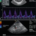
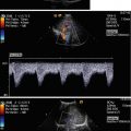
The normal aorta has clear defined smooth margins and tapers slightly below the level of the renal arteries. The abdominal aorta lies adjacent to the spine throughout its course, slightly to the left of the midline (Fig. 5.1). The inferior vena cava in contrast lies to the right of the midline and gradually moves away from the spine. In contrast to the inferior vena cava, the aorta has a straight course and hard pulsations (Fig. 5.1b). It gives origin to the coeliac trunk and the mesenteric arteries which can simultaneously be shown in longitudinal sections through the abdomen (Fig. 5.1a, b). Colour Doppler displays the artery red (Fig. 5.1c). Pulsed wave Doppler shows a laminar flow characterised by the typical window under the flow curve (Fig. 5.1d). The flow curve is triphasic with a systolic forward flow, an end-systolic negative flow and a very low diastolic forward flow (Fig. 5.1d). This characterises the abdominal aorta as a high-resistance vessel: The abdominal aorta perfuses the low-resistance abdominal organs on one hand and on the other hand, equally important, the high-resistance lower extremities. If flow measurements are performed, flow should be measured at the upper (or lower) end of the picture where the angle of incidence is lowest. The flow in these sections of the artery runs under an angle of 30–50° to the Doppler beam. Correct flow velocities or volume flow can only be calculated if angle corrections are performed.

Fig. 5.1
Descending aorta (DAO): longitudinal section through the abdomen. (a) 2d image through the upper abdomen in a premature infant: longitudinal image through the descending aorta (DAO). From the aorta the coeliac trunk (TC) and the superior mesenteric artery (SMA) originate. VH hepatic vein. (b) 2d image: The aorta runs straight through the abdomen just in front of the vertebral spine. AMS arteria mesenterica superior, DAO descending aorta, TC coeliac trunk, VMS vena mesenterica superior. (c) Colour-coded Doppler sonography of flow within the aorta and its abdominal branches. The flow in the descending aorta (DAO), the superior mesenteric artery (AMS) and the coeliac trunk (TC) is displayed red. (d, e) Spectral Doppler recording of the flow within the descending aorta. The picture shows a laminar triphasic flow profile with a systolic peak, a short end-systolic backflow and a low end-diastolic forward flow, characteristic of a high-resistance artery
The descending aorta can be shown on axial sections through the abdomen too (Fig. 5.2). Depending on the height of the section, the origin of different arteries such as the coeliac trunk, the superior mesenteric artery and both renal arteries can be shown. Below the umbilicus the abdominal aorta bifurcates and gives origin to both common iliac arteries (Fig. 5.3). They run in front of the common iliac vein (Fig. 5.3b). Both vessels can be shown in longitudinal sections too (Fig. 5.3c). Flow in the iliac arteries is laminar and triphasic, as peripheral resistance is high (Fig. 5.3d).


Fig. 5.2
(a) Transverse section through the upper abdominal aorta in the region of the origin of the coeliac artery (asterisk). DAO descending aorta, GB gall bladder, HA hepatic artery, IVC inferior vena cava, PV portal vein, SA splenic artery. (b) 2d expanded image of the coeliac trunk: HA hepatic artery, SA splenic artery, TC coeliac trunk. (c, d) Colour-coded Doppler sonography of the flow in the descending aorta (DAO) and coeliac trunk (CT): HA hepatic artery, SA splenic artery
Fig. 5.3
Greyscale sonography of the iliac vessels. (a) Transverse section through the lower abdomen in the region of the aortic bifurcation below the umbilicus. The image shows both common iliac arteries. (b) Transverse section through the right lower abdomen shows the common iliac artery (IA) and vein (IV). P iliopsoas muscle. (c) Longitudinal image of the common iliac artery (CIA) and vein (CIV).(d) Pulsed Doppler recording of the flow in the common iliac artery: laminar triphasic flow characteristic of a high-resistance flow
Doppler sonographic flow measurements in the descending aorta cannot be performed in transverse sections, as the angle of incidence approximates 90°. With colour-coded Doppler sonography, flow can be seen: If the transducer is slightly angled cranially, the artery is displayed red as the flow runs towards the transducer (Fig. 5.2b). If the transducer is slightly angled caudally, the artery is displayed blue as the flow runs away from the transducer.
The first abdominal branch which originates from the aorta is the coeliac trunk; the second is the mesenteric artery (Figs. 5.1, 5.2, 5.4 and 5.5a, b). Both arteries can be seen in longitudinal sections through the abdomen (Figs. 5.1a, b and 5.5a, b). The third arteries which arise laterally from the aorta below the superior mesenteric artery are the renal arteries (Fig. 5.14). In contrast to the coeliac trunk and the superior mesenteric artery, the renal arteries are best displayed in axial views below the origin of the superior mesenteric artery (Fig. 5.14). Below the umbilicus the abdominal aorta gives origin to both common iliac arteries which can be displayed as round pulsating structures laterally to the midline (Fig. 5.3a). In longitudinal sections, the iliac arteries are displayed as tubular structures (Fig. 5.3c).


Fig. 5.4
Schematic drawing of the truncus coeliacus and its tributaries. SA splenic artery, AMS superior mesenteric artery, GDA gastroduodenal artery, LHA left hepatic artery, LGA left gastric artery, RGA right gastric artery, RHA right hepatic artery
Fig. 5.5
(a) Longitudinal section through the origin of the coeliac artery. The coeliac trunk (CT) is the first vessel which originates from the descending aorta (DAO). Normally the branching of the artery into the hepatic artery (asterisk) or the splenic artery cannot be shown in longitudinal sections. SMA superior mesenteric artery. (b) Longitudinal section through the origin of the coeliac artery. DAO descending aorta, SMA superior mesenteric artery; 1 coeliac trunk, 2 common hepatic artery, 3 splenic artery. (c) Transverse section through the coeliac artery (asterisks). The coeliac trunk bifurcates in the hepatic artery (HA) to the patient’s right and the splenic artery (SA) to the patients left. DAO descending aorta; PV portal vein, IVC inferior vena cava. (d) Transverse section through the coeliac axis angled cranially. Cranially to the origin of the splenic artery (not displayed), the origin of the left gastric artery (GA) can be shown
5.1.2 Coeliac Artery
The coeliac artery, also called the coeliac trunk or the coeliac axis, is the first branch of the abdominal aorta (Fig. 5.4). It originates from the anterior surface of the aorta, between the diaphragmatic crura. It bifurcates after 0.5–2 cm from its origin into the common hepatic artery and the splenic artery, which can easily be visualised by ultrasound in transverse sections (Figs. 5.2a and 5.5c). The left gastric artery, which originates from the coeliac trunk too, usually cannot be visualised in infancy and childhood. In adolescents however the left gastric artery can be displayed (Fig. 5.5d).
The coeliac artery can be displayed either in sagittal (longitudinal) sections or in axial (transverse) sections through the upper abdomen (Figs. 5.1, 5.2 and 5.5a–c). The T-shaped bifurcation and its branches can best be shown in transverse planes (Figs. 5.2a,b and 5.5c). In longitudinal sections the branches of the artery are not well seen, although in rare cases branches of the coeliac trunk can be demonstrated (Fig. 5.5a, b).
Colour Doppler displays the flow red, as the blood stream is directed towards the transducer (Fig. 5.5a, b).

Fig. 5.6
(a) Colour Doppler of the flow in the descending aorta (DAO), the coeliac trunk (CT) and the superior mesenteric artery (SMA) (longitudinal section). (b) Colour flow in the coeliac artery (TC) in transverse section through the upper abdomen. DAO descending aorta, IVC inferior vena cava, PV portal vein, SA splenic artery, TC coeliac trunk. (c, d) Spectral Doppler of the flow in the coeliac artery in longitudinal (c) and transverse (d) section. The sample volume is placed just above the origin of the artery. The flow profile is monophasic with high diastolic amplitude due to low peripheral vascular resistance. (e) Flow measurement of the flow in the superior mesenteric artery shows systolic-diastolic forward flow with low diastolic amplitude due to high peripheral resistance
Pulsed wave Doppler shows a pulsatile flow displayed over the baseline. The flow profile is monophasic with a large amount of continuous forward flow throughout diastole (Fig. 5.6c–d). The monophasic flow is caused by the low peripheral resistance in the perfused organs of the upper abdomen (liver, spleen, pancreas). The splenic and hepatic branches show the same low-resistance flow pattern, as the peripheral resistance within the microcirculation of the liver and the spleen is low. Normal values for the flow velocities in the coeliac artery are helpful for the judgement of pathologic flow (Table 5.3).
The coeliac trunk is the ideal vessel for Doppler sonographic flow measurements in the abdomen:
1.
It is relatively large, even in premature babies.
2.
It has an ideal orientation from the aorta to the surface of the skin: Doppler sonographic flow measurements can be performed without angle correction, as the angle approximates zero.
3.
Flow alterations in other arteries, such as the cerebral arteries, may be caused by diseases of the brain or haemodynamic causes. To answer these questions, flow measurements in the coeliac axis are helpful. If similar changes are found in cerebral and abdominal arteries, a haemodynamic cause (congenital heart disease, shock etc.) is very probable and should be ruled out. If the flow changes are only found in the cerebral arteries, diseases of the central nervous system are more probable. If flow changes occur only in the coeliac trunk, abdominal causes should be ruled out (Table 5.3).
Table 5.3
Normal values of the flow velocities and the resistance indices in the coeliac trunk of prematurely born infants and healthy newborns
Premature infants | Newborns | |
|---|---|---|
V s | 72.9 ± 11.1 | 77.6 ± 20.8 |
V es | 31.6 ± 9.9 | 28.5 ± 7 |
V ed | 20.6 ± 7 | 15.3 ± 4.3 |
TAV | 19.9 ± 5 | / |
RI | 0.72 ± 0.10 | 0.79 ± 0.10 |
5.1.2.1 Variations
The branching pattern of the coeliac axis into the common hepatic artery, the splenic artery and the left gastric artery is quite constant, occurring in 93 % of all individuals (Zwiebel 2005). In less than 1 %, the coeliac artery and the superior mesenteric artery arise from the aorta as a common trunk. In these variations the common trunk splits into the coeliac artery and the superior mesenteric artery within several millimetres to 1 or 2 cm (Michels 1955, Ruzika and Rossi 1970).
5.1.3 Hepatic Artery
The common hepatic artery is the limb of the T-shaped coeliac axis that turns to the patient’s right (Figs. 5.2, 5.5 and 5.7). The artery runs a short distance at the superior border of the pancreatic head and gives rise to the gastroduodenal artery which can sometimes be seen at the anterosuperior border of the pancreatic head in older children. Beyond the gastroduodenal artery, the common hepatic artery becomes the proper hepatic artery which follows the portal vein to the porta hepatis. At the entrance to the liver, it divides into the left and right hepatic arteries, which penetrate into the liver. Due to anatomic rules, branches of the hepatic artery, portal vein and bile ducts always run together.

Fig. 5.7
(a) Transverse section through the upper abdomen shows the origin of the hepatic artery (AH) and the splenic artery (SA) from the coeliac trunk (asterisks). DAO descending aorta, GB gall bladder, IVC inferior vena cava. (b) Proper hepatic artery (HA) (transverse section through the liver) GB gall bladder, LPV left portal vein, RPV right portal vein. (c) Colour Doppler of the flow in the proper hepatic artery (HA) at the porta hepatis. Although the angle of incidence is bad, the flow can be shown. VP portal vein. (d) Colour Doppler of the flow in the hepatic vessels displayed in a transverse section through the liver. The transducer is slightly angled cranially. Due to low flow velocities, the portal vein (PV) is displayed in dark red colour. The flow velocities in the hepatic artery (HA) are much higher; the artery is therefore displayed in bright red. HV hepatic vein. (e) Spectral Doppler of the flow in the hepatic artery. Systolic-diastolic forward flow with high diastolic amplitude, characteristic of the low-resistance flow in the hepatic artery
The hepatic arteries can be visualised sonographically from an anterior abdominal approach. The common hepatic artery is most easily identified at its origin from the coeliac artery in transverse sections through the upper abdomen (Figs. 5.5c and 5.7a). Sometimes the origin of the artery can be displayed in longitudinal sections too (Fig. 5.5a, b).
The proper hepatic artery is seen near the porta hepatis that shows the portal vein in short or better long axes. The right and left hepatic arteries can be followed into the liver to a variable distance in children and adolescents (Fig. 5.7b). Due to the small size of the vessels in preterm babies, infants and toddlers, it is often not possible to display the branches of the hepatic artery on two-dimensional images. Using colour Doppler and high flow settings (low sensitivity and high PRF), the flow in the portal veins can be depressed and the high flow in the branches of the hepatic arteries can be seen adjacent to the portal vein branches (Fig. 5.7d). If higher sensitivity (lower PRF) is used and flow in the portal vein and the hepatic artery are simultaneously displayed, aliasing occurs in the hepatic artery (Fig. 5.7c).
Depending on the course of the common hepatic artery, the flow is displayed blue or red if colour-coded Doppler sonography is used (Fig. 5.7d). If the flow is slightly directed towards the transducer, the artery is displayed red; in the other cases (right hepatic artery), it is displayed blue. Flow in the left branch of the hepatic artery runs towards the transducer and is always displayed red (Fig. 5.7d); flow in the right branch of the artery runs away from the transducer and is therefore displayed blue.
As mentioned previously flow in the hepatic arteries is monophasic with high diastolic amplitude, due to low peripheral resistance (Fig. 5.7e).
5.1.3.1 Variations
The classic hepatic artery configuration is found in 72 % of all individuals (Taylor et al. 1985). Many other variations may occur. The most common are:
The common (4 %) or right hepatic artery (11 %) may arise from the superior mesenteric artery.
The left hepatic artery may arise from the left gastric artery (10 %) (Michels 1955).
5.1.4 Splenic Artery
The splenic artery is the left limb of the coeliac artery. It runs along the posterior superior margin of the pancreatic body and tail before it turns anteriorly to the spleen (Figs. 5.2, 5.5 and 5.7). At the splenic hilus it splits into several branches.
Along its way the splenic artery gives rise to several pancreatic branches, short gastric branches and the left gastroepiploic artery. None of these vessels can be shown sonographically in infancy and childhood.
The splenic artery can be displayed by ultrasound along its complete course. The best images are transverse sections through the upper abdomen which show the artery at the superior border of the pancreas (Figs. 5.7a and 5.8a). The middle portion of the artery and vein can be shown in longitudinal sections too if the transducer is slightly angled to the left side (Fig. 5.8b). The artery always runs together with the splenic vein (Fig. 5.8b). Using 2d images both vessels cannot be distinguished for sure. Usually the artery is the cranially localised vessel. The different Doppler techniques (colour or spectral Doppler) however can easily identify arteries and veins.

Fig. 5.8
2d imaging of the splenic artery and vein. (a) Transverse section through the left upper abdomen in a 13-year-old boy. The image shows the splenic artery (SA) as it curves around the posterior margin of the pancreas (P). CT coeliac trunk, DAO descending aorta, L liver. (b) Sagittal section through the upper abdomen in a 13-year-old boy. The transducer is slightly angled to the left. Both splenic vessels can be seen behind the pancreas (P). The splenic artery (SA) lies cranially and is smaller than the caudally located splenic vein (SV). L liver
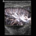
The distal branches of the vein can best be shown in transverse or coronal sections through the splenic hilus from a left approach (Fig. 5.9).

Fig. 5.9
Transverse section through left upper abdomen in the region of the splenic hilus shows the splenic artery (markers) as it curves around the tail of the pancreas (P). (a) 2d transverse image through the upper abdomen shows a vessel entering the splenic hilus. P pancreas, K right kidney, S spleen. (b) Colour Doppler of the vessels at the splenic hilus. The arteries are displayed red, the veins blue
Using colour-coded Doppler sonography, the flow in the artery behind the pancreas can be displayed blue as the flow is directed away from the transducer (Fig. 5.10a, b). When the artery has passed the tail of the pancreas and turns towards the splenic hilus, it is displayed red (Fig. 5.10f).

Fig. 5.10
Colour Doppler of the flow in the splenic artery. (a) Transverse section through the upper abdomen and the pancreatic body. The flow in the splenic artery (SA) is displayed blue. AO aorta, IVC inferior vena cava, PV portal vein, superior mesenteric artery (asterisk). (b) Spectral Doppler of the flow in the splenic artery behind the body of the pancreas. The sample volume is placed in the splenic artery which is displayed blue. The flow spectrum shows a monophasic flow profile with high diastolic amplitude and a broad flow spectrum. The flow is displayed below the baseline as it is directed away from the transducer. (c) Colour flow imaging of the splenic vessels in a sagittal section through the upper abdomen of a 13-year-old boy. The transducer is slightly angled to the left. The flow in the cranially located splenic artery (SA) is displayed blue, whereas the caudally located splenic vein (SV) is displayed red. (d) Spectral Doppler of the flow in splenic artery shows a systolic-diastolic flow displayed below the baseline. (e, f) Spectral Doppler of the flow in the splenic artery at the splenic hilus from a transverse lateral approach. The artery is displayed in a mosaic pattern indicating high flow. The flow curve is monophasic with high diastolic forward flow, due to low peripheral resistance
5.1.5 Left Gastric Artery
The left gastric artery is the third branch of the coeliac artery. The left gastric artery is the smallest of the branches. The small artery cannot routinely be shown in infants and young children.
In older individuals and adolescents, the left gastric artery can be displayed cranially of the pancreas. It turns to the left and can be seen at the upper border margin of the stomach in the region of the gastroesophageal communication (Fig. 5.11a). Beside the artery the left gastric vein can sometimes be displayed (Fig. 5.11a). Colour Doppler displays the flow in the artery blue (Fig. 5.11b). Pulsed Doppler reveals as systolic-diastolic forward flow with low diastolic amplitude due to intermediate peripheral vascular resistance (Fig. 5.11c).

Fig. 5.11
(a) Left gastric artery in a transverse section through the upper abdomen (the transducer is slightly angled cranially and rotated to the left). Beside the left gastric artery (AG) the left gastric vein (VG) is displayed. AO aorta. (b) Colour Doppler of the flow in the left gastric artery (GA) which originates from the coeliac trunk (CT). PV portal vein. (c) Spectral Doppler of the flow in the left gastric artery: systolic-diastolic forward flow with low diastolic amplitude due to higher resistance during fasting
5.1.6 Superior Mesenteric Artery (SMA)
The superior mesenteric artery arises as the second branch from the anterior surface of the aorta, immediately distal to the coeliac trunk (Figs. 5.1a, 5.4, 5.5a, b and 5.12a). The SMA consists of a short, anteriorly directed segment and a longer inferiorly directed segment that ends in the region of the ileocaecal valve (Fig. 5.12a). SMA branches supply the jejunum, ileum, caecum and ascending colon, as well as the proximal two-thirds of the transverse colon and portions of the duodenum and pancreatic head.

Fig. 5.12
(a) Longitudinal section through the upper abdomen and the superior mesenteric artery (SMA). The artery parallels the descending aorta (DAO). In front of the SMA the superior mesenteric vein can be displayed. (b) Transverse section through the upper abdomen and the superior mesenteric artery (SMA). The SMA is surrounded by a typical halo of echogenic fat. SV splenic vein, VCI inferior vena cava
The artery can be displayed in longitudinal and transverse sections through the upper abdomen (Figs. 5.1a, b, 5.5a and 5.12a, b). In transverse planes the artery is displayed between the splenic vein anteriorly and the aorta posteriorly. The SMA is surrounded by an echogenic halo of fat (Fig. 5.12b). The SMA is an important anatomic landmark for scanning of upper abdominal vessels. The SMA lies to the left of the superior mesenteric vein. It lies behind the body of the pancreas and the splenic vein. The left renal vein lies posterior to the SMA. It runs between the aorta and the SMA.
Colour Doppler reveals the flow in the SMA. It runs best in longitudinal sections. The artery is displayed red as the flow is directed towards the transducer (Figs. 5.1b and 5.6a).
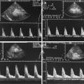
Measurements of flow in the SMA are best performed in the proximal section of the SMA where the angle of incidence is low. The angle of incidence in the section where the artery parallels the aorta approximates 90°. Flow measurements cannot be performed in this section. The flow in the SMA depends highly on the fasting status of the patient. In contrast to the flow in the coeliac artery, a high-resistance flow pattern with a sharp systolic peak and low diastolic amplitude can be found in the fasting patient (Fig. 5.13a, b). Sixty minutes after a meal, the diastolic amplitude increases to a low-resistance flow profile with a high diastolic amplitude similar to the flow in the coeliac trunk (Table 5.4).

Fig. 5.13
(a, b) Doppler sonographic flow measurement in the coeliac trunk (a) and the superior mesenteric artery (SMA). The image shows a significant higher diastolic forward flow in the coeliac trunk than in the superior mesenteric artery due to the fasting status of the patient
Table 5.5
Prothrombotic risk factors
Antithrombin deficiency |
Protein C deficiency |
Protein S deficiency |
Plasminogen deficiency |
Factor V Leiden mutation |
Prothrombin gen 202110A |
Dysfibrinogenaemia |
Antiphospholipid antibodies |
Hyperhomocystinaemia |
Increased lipoprotein a |
5.1.7 Renal Arteries
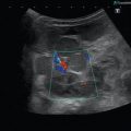
The renal arteries arise from the aorta, slightly below the SMA. The right renal artery arises from the anterolateral aspect of the aorta before it passes under the inferior vena cava and then enters the right renal hilus (Fig. 5.14a). The left renal artery originates from the left lateral or posterolateral aspect of the aorta before it follows a posterolateral course to the left hilus (Fig. 5.30c).

Fig. 5.14


(a, b) Transverse section through the origin of the right renal artery (RA). The RA can be displayed from an anterior approach behind the inferior vena cava (IVC). DAO descending aorta, PV portal vein. (c) Longitudinal section through the inferior vena cava (IVC). Behind the IVC the right renal artery (RA) which compresses the posterior wall of the IVC can be displayed. HA hepatic artery, VP portal vein
Related posts:
Stay updated, free articles. Join our Telegram channel
Full access? Get Clinical Tree
