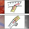
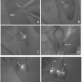
Fig. 41.1
Dynamic indocyanine green lymphography
41.2 Dynamic ICG Lymphography
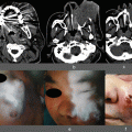
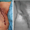
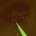
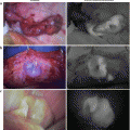
Dynamic ICG lymphography consists of 2 phase observations: early transient phase for evaluation of lymph pump function, and late plateau phase for evaluation of lymph circulation and for navigation of lymphatic surgery [4, 5, 8, 11–13]. Dynamic ICG lymphography is performed as follows: An examinee is kept still for 15 min, and 0.05–0.2 ml of ICG (Diagnogreen 0.25 %; Daiichi Pharmaceutical, Tokyo, Japan) is subdermally injected (at the 2nd web space for extremity and genital lymphedema, and at the glabella and the philtrum for facial lymphedema). Immediately after ICG injection, fluorescent lymph flow images are obtained using an infrared camera system (transient phase). Examinees are kept still in supine position during lymph pump function measurement. After completion of the measurement (5 min after injection), examinees are allowed to move freely [12, 13].
Approximately 12–18 h after ICG injection, ICG movement reaches a plateau (plateau phase). In a plateau phase, lymph circulation is assessed based on ICG lymphography findings as mentioned in Sect. 43.2.2, which allows pathophysiological severity staging and pre- and intraoperative navigation for lymphatic surgery [3–11, 14–16]. For more rapid and convenient assessment, patients are better to be instructed to move their extremity rigorously. When patients move their extremity rigorously, ICG can reach a plateau 2 h after ICG injection. Plateau phase continues until 72 h after ICG injection; evaluation of abnormal lymph circulation is possible between 2 and 72 h after ICG injection.
41.2.1 Assessment of Lymph Pump Function
A major strength of ICG lymphography is real-time evaluation. Lymph pump function can be easily and directly measured by observing ICG movement at an early transient phase. Several methods have been reported to evaluate lymph transportation capacity, and ICG velocity is the most practical one; unlike other measurements that sometimes require 1 h for lymphedema evaluation, measurement of ICG velocity can be completed within 5 min after ICG injection [12, 13]. The distance between the injection point and the farthest point where ICG is observed is measured 5 min after ICG injection. ICG velocity is calculated as follows:
 When ICG reaches the groin/axilla in extremity lymphedema cases within 5 min, ICG velocity is calculated by dividing the distance between the groin/axilla and the injection point by the time required for transition. ICG velocity decreases with lymphedema progression and increases after successful treatments. As ICG velocity is a quantitative measurement, it is quite easy to evaluate lymph transportation capacity before and after treatments.

41.2.2 Assessment of Lymph Circulation
With progression of obstructive lymphedema, ICG lymphography pattern changes from normal linear pattern to abnormal dermal backflow (DB) patterns. DB patterns include splash (mild DB), stardust (moderate DB), and diffuse (severe DB) patterns (Fig. 41.2) [3]. Lymph flow is obstructed after dissection and/or radiation of lymph nodes, which leads to lymphatic hypertension, dilatation of lymphatic vessels, lymphatic valve insufficiency, lymphosclerosis, and lymph backflow. Lymph backflow is visualized as DB patterns on ICG lymphography [3–7, 17, 18]. Splash pattern represents dilated superficial lymphatic precollectors and capillaries that can work as collateral lymph pathways. Lymph extravasation takes place when the collateral fails to compensate lymph overload, which is visualized as spots on ICG lymphography, stardust pattern. With progression of lymph extravasation, the number of spots on ICG lymphography increases the point where spots cannot be distinguished from each other, diffuse pattern. In obstructive lymphedema, DB patterns usually extend from proximal to distal region. Assessment of abnormal lymph circulation using ICG lymphography allows pathophysiological severity staging and prediction of lymphatic vessels before lymphatic surgery.
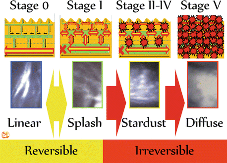

Fig. 41.2
Indocyanine green lymphography findings and lymph circulation
41.2.2.1 Pathophysiological Severity Staging Systems for Secondary Lymphedema (Dermal Backflow Stages)
Pathophysiological severity staging system for obstructive lymphedema, DB stage, is determined based on ICG lymphography findings. There are 4 DB stages: leg DB (LDB) stage for leg lymphedema, arm DB (ADB) stage for arm lymphedema, genital DB (GDB) stage for genital and lower abdominal lymphedema, and facial DB (FDB) stage for head and neck lymphedema [4–7, 19].
LDB stage is a severity staging system for obstructive lower extremity lymphedema (Fig. 41.3) [3, 4, 11, 20]. In LDB stage 0, no DB pattern is observed. In LDB stage I, splash pattern is observed around the groin. In LDB stage II through V, stardust pattern is observed. In LDB stage II, stardust pattern is limited proximal to the patella. In LDB stage III, stardust pattern exceeds the patella. In LDB stage IV, stardust pattern is detected throughout the lower extremity. In LDB stage V, diffuse pattern is observed with background of stardust pattern.
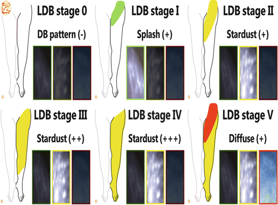

Fig. 41.3
Leg dermal backflow (LDB) stage for pathophysiological severity staging of leg lymphedema using indocyanine green lymphography
ADB stage is a severity staging system for obstructive arm lymphedema (Fig. 41.4) [5, 11, 20]. In ADB stage 0, no DB pattern is observed. In ADB stage I, splash pattern is observed around the axilla. In ADB stage II through V, stardust pattern is observed. In ADB stage II, stardust pattern is limited proximal to the olecranon. In ADB stage III, stardust pattern exceeds the olecranon. In ADB stage IV, stardust pattern is detected throughout the upper extremity. In ADB stage V, diffuse pattern is observed with background of stardust pattern.
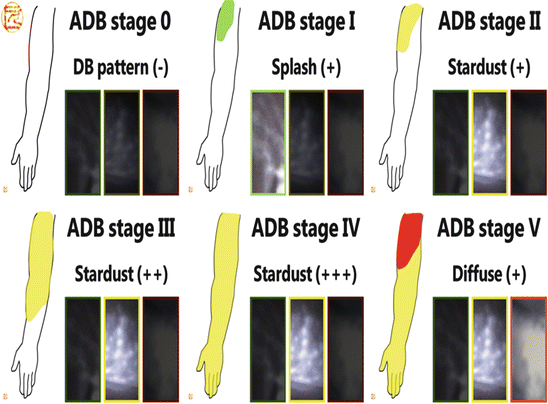

Fig. 41.4
Arm dermal backflow (ADB) stage for pathophysiological severity staging of arm lymphedema using indocyanine green lymphography
GDB stage is a severity staging system for obstructive lower abdominal and genital lymphedema [6, 11, 20, 21]. In GDB stage 0, no DB pattern is observed. In GDB stage I, splash pattern is observed around the groin and/or lower abdominal region. In GDB stage II, stardust pattern is seen around the groin and/or lower abdominal region. In GDB stage III, stardust pattern is observed around the groin, the lower abdominal, and the genital region. In GDB stage IV, diffuse pattern is observed with background of stardust pattern.
FDB stage is a severity staging system for obstructive head and neck lymphedema [7, 11, 20]. In FDB stage 0, no DB pattern is observed. In FDB stage I, splash pattern is observed around the neck. In FDB stage II through V, stardust pattern is observed. In FDB stage II, stardust pattern is observed in the neck. In FDB stage III, stardust pattern is seen in the face and neck. In FDB stage IV, stardust pattern is detected throughout the head and neck region. In ADB stage V, diffuse pattern is observed with background of stardust pattern.
41.2.2.2 ICG Lymphography Classification for Primary Lymphedema
Since primary lymphedema has a wide variety of etiologies, it is important to visualize abnormalities in lymphatic systems for evaluation and diagnosis. Although ICG lymphography can visualize only superficial lymph flows, ICG lymphography has been reported to be useful to evaluate and classify primary lymphedema [22]. The ICG lymphography primary lymphedema classification includes four patterns: proximal DB (PDB), distal DB (DDB), less enhancement (LE), and no enhancement (NE) patterns (Fig. 41.5).
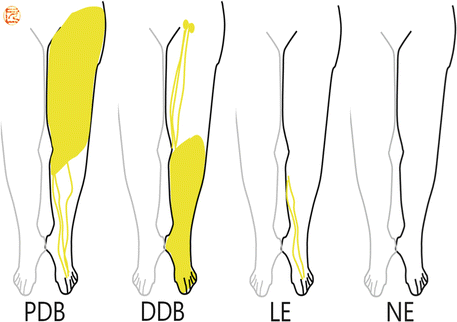

Fig. 41.5
Indocyanine green lymphography classification for primary lymphedema. Proximal dermal backflow (PDB) pattern, distal dermal backflow (DDB) pattern, less enhancement (LE) pattern, and no enhancement (NE) pattern
In PDB pattern, DB pattern extends from proximal to distal region like DB stages for obstructive lymphedema. Patients with PDB pattern should undergo work-up to rule out malignancy because the findings are very similar to that of cancer-related lymphedema. Lymphatic malformation in the trunk is suspected. Therapeutic strategy is also similar to that of cancer-related obstructive lymphedema. Earlier interventions work better to improve lymph circulation [23].
In DDB pattern, DB pattern is observed in the distal part but not in the proximal part, and the remaining region shows linear pattern or no enhanced images. Localized distal malformation or lymphatic valve malformation is suspected. Patients with DDB pattern usually have past history of cellulitis, and lymphatic vessels are likely to be affected by inflammation.
In LE pattern, linear pattern is observed only in the distal part, and the remaining proximal part shows no enhanced image; no DB pattern is detected. Relative hypoplasia of superficial lymphatic system is suspected, and aging-related pump dysfunction may affect development of symptomatic lymphedema. Patients with LE pattern usually find themselves prone to physiologic temporary leg swelling before onset of pathologic and progressive leg edema.
Stay updated, free articles. Join our Telegram channel

Full access? Get Clinical Tree


