Chapter 77 This chapter covers congenital anomalies of the aorta and the pulmonary arteries, with an emphasis on anomalies that produce clinical symptoms of airway and esophageal obstructions. Anomalies of the thoracic great arteries can be broadly classified into anomalous origins, anomalous connections, obstructions, and structural anomalies of the aortic arch. Important clinical entities under each category are listed in Box 77-1. This chapter will focus on structural anomalies of the aortic arch and pulmonary sling. Overview: Anomalies of the aortic arch and the cervical vessels are relatively common, with a prevalence estimated at 0.5% to 3%, depending on the inclusion criteria. Most variations, such as a left aortic arch with an aberrant right subclavian artery, common origin of the left common carotid and right innominate arteries (bovine arch), and ectopic origin of the left vertebral artery from the aortic arch, are of little or no clinical consequence. “Vascular ring” is a term that refers to encirclement of the trachea and esophagus caused by the abnormal embryologic development of the aortic arch. The principal structural components responsible for this encirclement are derived from the aortic arch or arches, subclavian artery, circumflex aortic segment, ductus arteriosus, or ligamentum arteriosum. Structural anomalies of the aortic arch can be understood as the abnormal divisions in the totipotential arch, a theoretical construct proposed by Jesse Edwards (Fig. 77-1).1 Only a small subset of aortic arch developmental anomalies leads to vascular rings, accounting for less than 1% of all congenital cardiovascular defects. Although about a dozen different types of vascular rings exist, double aortic arches and right aortic arch with left ligamentum arteriosum account for 90% of cases.2 Figure 77-1 Ventral view of Dr. Jesse Edward’s “totipotential arch” or hypothetic double aortic arch and bilateral ducti arteriosi. Chromosome 22q11 deletion has been reported in 24% of patients with isolated arch anomalies.3,4 This deletion was first identified in persons with DiGeorge syndrome, which consists of various degrees of immunodeficiency, thymic hypoplasia or aplasia, hypoparathyroidism, outflow tract cardiac defects, and dysmorphic appearance. Chromosome 22q11 deletion is now recognized as a major factor in many congenital heart defects. For example, it is detected in up to 50% of patients born with interrupted aortic arch or truncus arteriosus. Testing for this chromosomal abnormality can be performed using fluorescence in situ hybridization. Clinical Manifestations: The severity of symptoms and the age of onset depend on the extent of the compression about the esophagus and trachea.5 Because different ring arrangements have different constrictive effects, not all vascular rings produce the same degree of symptoms; in fact, some rings produce no symptoms. Conversely, symptomatic vascular compression of the trachea and esophagus does not require a complete ring, as can be seen in an anomalously placed or aneurysmal innominate artery or a retroesophageal subclavian artery. Most cases of vascular ring, if they are symptomatic, present during infancy or early childhood. Imaging: Chest radiography may show tracheal compression by a vascular ring, but by itself a chest radiograph cannot confirm or exclude a vascular ring. Because a vascular ring is more likely in the presence of a right aortic arch, symptoms of tracheal compression in the presence of a right aortic arch should raise the possibility of a vascular ring. In patients presenting with nonspecific symptoms, barium esophagography is a useful first test.6 A normal barium esophagram usually excludes a clinically significant vascular ring. Classic S-shaped indentations in the frontal projection of an esophagram are highly suggestive of double aortic arches. A posterior vascular indentation may or may not be a vascular ring but is more likely in the presence of a right aortic arch. An esophagram may reveal other causes of a patient’s symptoms, such as gastroesophageal reflux, aspiration, or tracheoesophageal fistula. Mediastinal ultrasonography or echocardiography with gray scale and color Doppler imaging may visualize the vascular ring directly in neonates and infants because these patients have excellent sonographic windows.7 Echocardiography generally is not useful in older children or adolescents. Moreover, because ultrasound is primarily a two-dimensional imaging method, connecting tortuous vascular structures can be difficult, especially when ligamentous or interrupted vascular segments are present. The presence of a vascular ring has been diagnosed successfully in utero with fetal ultrasonography.8 Patients with severe symptoms or an abnormal results of an esophagram, chest radiograph, or mediastinal ultrasonography should undergo angiography to confirm the vascular abnormalities and to gather information for surgical planning. Conventional catheter angiography has been replaced by first-pass, contrast-enhanced computed tomographic angiography (CTA) or magnetic resonance angiography (MRA). Both modalities can visualize the aortic arch and the cervical arteries well. MRA is usually preferred because it does not subject the patient to ionizing radiation.9,10 However, in cases in which the airways and the lungs must be evaluated together with the vascular anomaly, CTA can accomplish both in a single scan and may be a better choice. Treatment: The definitive treatment is surgical relief of the obstruction.11 A double aortic arch is repaired by dividing the nondominant arch between its last cervical artery and the point where the nondominant arch joins the descending aorta. If the ductus arteriosus or the ligamentum arteriosum forms a border of the ring, it is ligated to relieve the constriction. A thoracotomy is performed at the side of the planned ligation. Persistent stenosis or tracheomalacia may develop in the constricted trachea, requiring additional repair. Double aortic arches can be categorized as bilaterally patent or as atretic in a portion of one of the two arches, usually the left. Bilaterally patent or complete double aortic arches represent persistence of both the right and the left embryologic fourth aortic arches. Two vessels arise from the ascending aorta and course dorsally, one on each side of the trachea and esophagus, to join posteriorly in a left descending aorta in 80% of cases. In this arrangement, the left arch is usually anterior and the right arch is posterior. In 20% of cases, the descending aorta lies on the right and the posterior-anterior relationship of the double aortic arches is reversed. The larger aortic arch is the dominant arch, and in 73% of cases, the right arch is dominant. The right arch normally is situated higher than the left arch, as can be seen in a typical esophagram, where the right arch indents the esophagus higher than does the left arch (Fig. 77-2). Double aortic arches usually are found without associated cardiac anomalies. Figure 77-2 Bilaterally patent double aortic arches in a 7-year-old boy.
Congenital Anomalies of the Thoracic Great Arteries
Vascular Rings
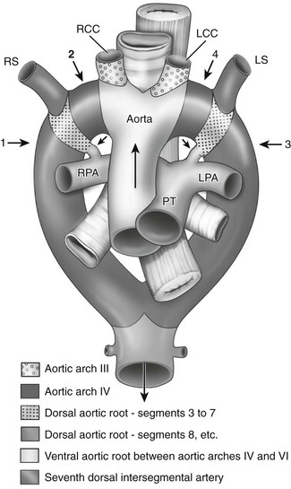
The numbered arrows point to the four key locations where regression occurs in various anomalies. Arrow 1 indicates the eighth segment of the right dorsal aortic root; arrow 2, the right fourth arch; and arrows 3 and 4, the corresponding two positions on the left. The shortest black arrows point to the ducti arteriosi bilaterally, and the longest black arrow indicates the direction of blood flow. LCC, Left subclavian artery; LPA, left pulmonary artery; LS, left subclavian artery; PT, pulmonary trunk; RCC, right common carotid artery; RPA, right pulmonary artery; RS, right subclavian artery. (From Stewart JR, Kincaid OW, Edwards JE: An atlas of vascular rings and related malformations of the aortic arch system, Springfield, IL, 1964, Charles C Thomas.)
Special Considerations
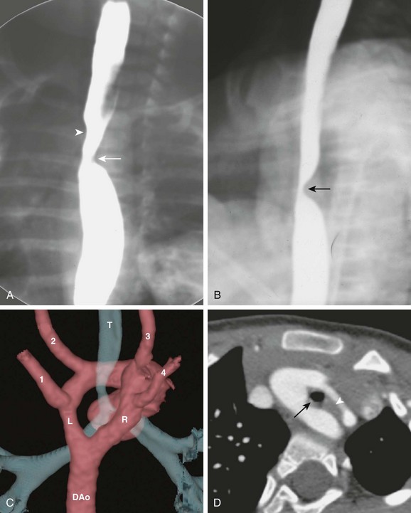
A, The frontal view of a barium esophagram shows bilateral indentations of the barium column. The right arch indention (arrowhead) is higher than the left (arrow). B, The lateral view of a barium esophagram shows a posterior indentation (arrow) corresponding to the posterior right arch. C, A volume-rendered image from a computed tomography (CT) angiogram shows the aorta as seen from the back. A dominant right arch (R) and a nondominant left arch (L) are present, both draining into the descending aorta (DAo). Four cervical arteries arise from the arch: left subclavian artery (1), left common carotid artery (2), right common carotid artery (3), and right subclavian artery (4). The ring encloses the trachea (T), causing tracheal narrowing just above the carina. D, An axial CT scan shows a complete double aortic arch constricting the trachea (black arrowRelated posts:
![]()
Stay updated, free articles. Join our Telegram channel

Full access? Get Clinical Tree


Congenital Anomalies of the Thoracic Great Arteries


