(1)
Department of Radiology, Saitama Medical University, Moroyama, Saitama, Japan
Abstract
The lesion has a well-defined border, and its wall is lined by spindle shaped cells.
12.1 Intra-articular Ganglion
The lesion has a well-defined border, and its wall is lined by spindle shaped cells.
Contains clear fluid similar to synovial fluid or mucoid material.
Commonly has multiple cystic chambers due to the presence of septa.
May cause pain and disorder of knee flexion and extension.
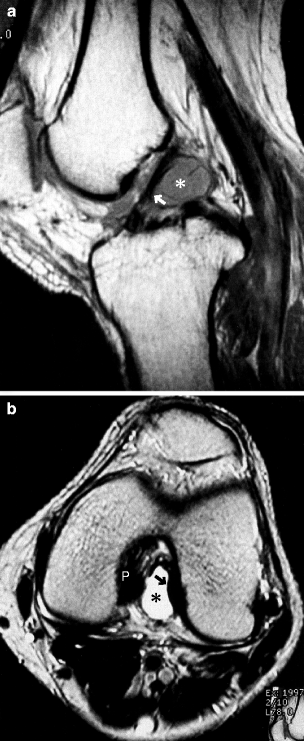
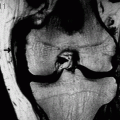
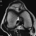
Fig. 12.1
ACL ganglion. A man in his 30s. (a) PDWI and (b) axial T2WI. There is an ACL ganglion (*) which is septated and compresses the ACL from below (arrow). P PCL
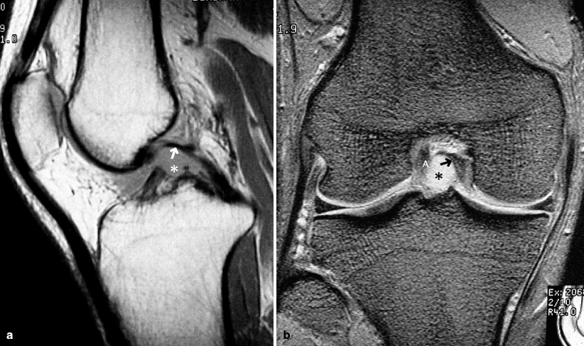
Fig. 12.2
PCL ganglion. A man in his 40s. (a) PDWI and (b) coronal T2*WI. There is a PCL ganglion (*). PCL shows an arc-like shape due to compression by the PCL ganglion (arrow). A ACL
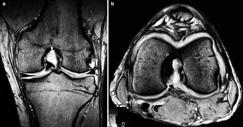
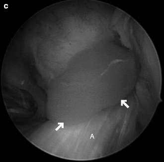
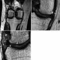
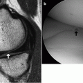
Fig. 12.3
ACL and PCL ganglion. A man in his 40s. (a) Coronal T2*WI, (b) axial T2*WI, and (c) arthroscopic image. There is a cystic lesion (arrow) sandwiched between ACL (A) and PCL (P), both of which are compressed by this lesion. Arthroscopic puncture of the cystic lesions revealed it was filled with yellow jellylike material
References
Bui-Mansfield LT, Youngberg RA. Intraarticular ganglia of the knee: prevalence, presentation, etiology and management. AJR. 1997;168:123–7.
Marra MD, Crema MD, Chung M, et al. MRI features of cystic lesions around the knee. Knee. 2008;15:423–38.
12.1.1 Ganglia and Bursae
A ganglion is a benign, unilocular, or multilocular cystic mass containing clear and highly viscous fluid within a dense fibrous connective tissue wall without a synovial lining. It does not communicate with the joint capsule. The pathogenesis of ganglia remains controversial. Proposed theories include mucoid cystic degeneration in a collagenous structure near areas under continuous stress and herniation of synovial tissue.
A bursa is lined by synovium and contains synovial fluid, usually representing normal physiologic fluid accumulation. Numerous bursae are present around the knee and have names according their anatomical location.
Ganglia and fluid-filled bursae are commonly seen around the joint and tendon sheath. They are often incidentally found on MRI in asymptomatic persons, but can cause pain and swelling. Ganglia tend to be multilocular, but differentiating between these two entities may be difficult at times on the basis of MRI alone.
12.2 Meniscal Cyst
It is a focal collection of synovial fluid located within or adjacent to the meniscus.
Parameniscal cysts are thought to form when there is fluid extravasation through a meniscal tear into the parameniscal soft tissue.
Large meniscal cysts may protrude laterally and may become palpable at the level of knee joint space as a subcutaneous mass (especially on the lateral side of the knee). Meniscal cysts may cause pain, tenderness, and swelling. Medial meniscal cysts are more likely to be painless.
Prevalence of the lateral meniscal cysts is 3–4 times higher than that of the medial meniscal cysts (Fig. 12.4). Meniscal cysts are particularly common around the anterior horn.
Medical meniscal cysts tend to enlarge into the posterior direction (Fig. 12.5).
Because the bond between the superficial layer of the MCL and the joint capsule is strong, it is rare for a cystic lesion to form at this location (see Fig. 5.3). However, in the event that fluid accumulates here, it may cause symptoms.
Meniscal cysts can be treated by surgical excision, but to prevent recurrence, meniscectomy may be necessary if the meniscal tear is present (Fig. 12.6).
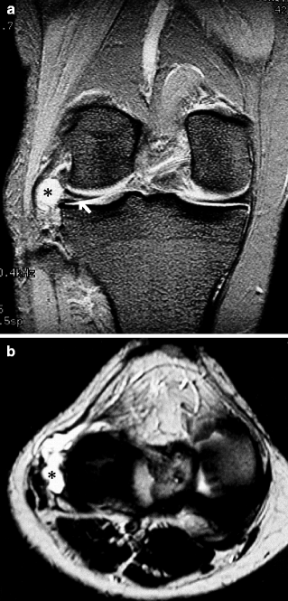
Fig. 12.4
Lateral meniscal cyst. A woman in her 40s. (a) Coronal T2*WI and (b) axial T2WI. There is a degenerative horizontal tear of the middle segment of the lateral meniscus (arrow) and a cystic lesion that is continuous with the tear (*)
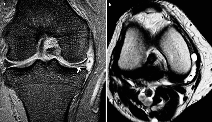
Fig. 12.5
Medial meniscal cyst. A woman in her 50s. (a) Coronal T2*WI and (b) axial T2WI. There is a degenerative horizontal tear of the medial meniscus (arrow) and a cystic lesion that is continuous with the tear and extends posteriorly (*)
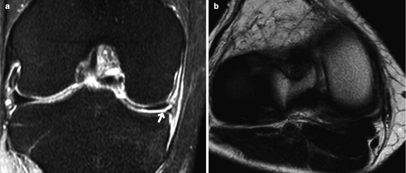
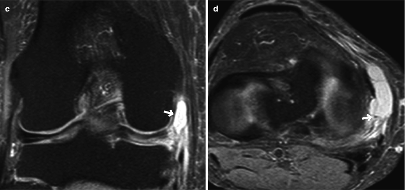
Fig. 12.6
Medial meniscal cyst that occurred 2 years after meniscal tear. A woman in her 40s. (a) Coronal T2*WI and (b) axial T2WI at the time of meniscal tear. (c, d) Respective sequences taken 2 years later. A medial meniscal tear is noted (arrow, a), but no cysts are seen. Two years later, there is a cystic lesion connected to the medial meniscal tear (arrows, c, d). This lesion was palpable
References
Jansen DL, Peterfy CG, Forbus JR, et al. Cystic lesions around the knee joint. MR imaging findings. AJR. 1994;163:155–61.
Tschirch FTC, Schmid MR, Pfirrmann CWA, Romero J, Hodler J, Zanetti M. Prevalence and size of meniscal cysts, ganglionic cysts, synovial cysts of the popliteal space, fluid-filled bursae, and other fluid collections in asymptomatic knees on MR imaging. AJR. 2003;180:1431–6.
12.3 Popliteal Cyst (Baker’s Cyst)
Popliteal cysts are not true cysts and represent fluid accumulation in the semimembranosus-medial gastrocnemius bursa (Figs. 12.7 and 12.8).
It commonly communicates with the joint capsule.
Most commonly seen cystic lesion in the whole body and the knee (about 40%).
On T2-weighted MRI, it shows homogeneous hyperintensity, but rarely it may appear heterogeneous if it contains hemorrhagic components or debris.
Increased intra-articular pressure due to joint effusion or other factors (e.g., meniscal tear, ACL tear, inflammatory arthritis) causes the extravasation of joint fluid through the posteromedial joint capsule posteriorly into the bursa, leading to gradual formation of an enlarging popliteal cyst.
It has a teardrop shape between the medial head of gastrocnemius and the semimembranosus tendon.
Rarely seen in children and becomes more common as the age increases.
Commonly painless if the size is less than 30 mm.
Rarely it can rupture (Fig. 12.9), causing extravasation of fluid into muscle interstitium and symptoms that are similar to those of thrombophlebitis.

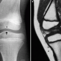
Fig. 12.7
Popliteal cyst. A man in her 20s. (a) Posterior view of the knee, (b) lateral radiograph, (c) axial T2WI, and (d) FS T2*WI. A swelling in the popliteal fossa (arrow


Related posts:
Stay updated, free articles. Join our Telegram channel
Full access? Get Clinical Tree
