George M. Bridgeforth and John Cherf
A 87-year-old man presents with left knee swelling and pain, which he has had for several months.
CLINICAL POINTS
- Swelling is often characteristic.
- Morning aching and stiffness.
- Older people are often affected.
Clinical Presentation
The clinical hallmark of degenerative joint disease, or osteoarthritis (OA), is hypertrophy with decreased range of motion. Mild swelling is common, and stiffness and soreness may be present. Patients sometimes describe the pain as an intense ache, and they may also complain that their knees creak or even “lock.” Degenerative joint disease has a predilection for the knees, hips, spine, and the small joints of the hands. The presence of other joint deformities characteristic of OA, such as Heberden nodes (hypertrophy of the distal interphalangeal joints) in some of the fingers, may be a helpful associated finding. The most common form of arthritis, degenerative joint disease, is the leading cause of chronic disability in the United States. It predominantly affects older individuals. The mechanism of injury is a degenerative process, and the condition develops our time.
The historical presence of locking or clicking inside of the knee is suspicious for a meniscus tear. However, locking may be caused by loose bodies or torn cartilage. The medial meniscus bears a greater load (i.e., more of the body weight), and a medial meniscus tear is more common than a lateral meniscus tear. Moreover, the medial meniscus is less mobile because it is firmly attached to the medial collateral ligaments and therefore more prone to damage from twisting injuries. Elderly patients with degenerative arthritis after develop a degenerative medial meniscus tear.
The presence of joint “mice” inside the knee may present floating loose bodies. This condition is characterized by the separation of small osteochondral body (i.e., small, usually rounded section of bone and cartilage). On a radiograph, it appears as a very small saucer-shaped defect inside the knee.
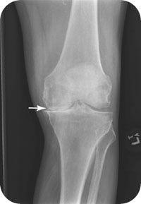
As previously stated, OA may be associated with mild to moderate swelling (Fig. 17.1). If the affected joint appears acutely warm, swollen, and tender, a diagnostic aspiration should be performed immediately. The differential diagnosis includes acute cellulitis (a neighboring open wound may serve as a portal for infection), gonorrhea, gout, rheumatoid arthritis, and psoriatic arthritis. OA is generally a bilateral condition; septic joints are usually unilateral. Treatment is directed at the underlying cause.
PATIENT ASSESSMENT 
- Limited range of motion
- Mild swelling and stiffness
- Pain, especially with weight-bearing
- Joint hypertrophy in later stages
Radiographic Evaluation
Radiographs that should be ordered include anteroposterior (AP), lateral, and oblique views (two). Two oblique views are used in a full trauma series to evaluate for condylar fractures of the tibial plateau. Optional views include the following:
- Sunrise view: used to check for subtle fractures and dislocations
- Merchant view: used to check for subtle fractures and dislocations. The preferred choice of many specialists, the Merchant view is superior to a sunrise view when checking for subtle fractures and dislocations.
- Cross-table lateral view: used as part of a trauma series. It is particularly useful when the patient has trouble bending the knee fully. The presence of a lipohemarthrosis indicates either an intra-articular fracture or derangement (i.e., cruciate ligament tear).
- Intercondylar view: used to better visualize the tibia spine. It is used to detect fractures of the median eminence (tibia spine).
NOT TO BE MISSED 
- Knee contusion or fracture
- Rheumatoid arthritis (usually bilateral)
- Gout (unilateral but may be bilateral)
- Psoriatic arthritis (oligoarticular)
- Septic joint (unilateral)
Related posts:


Stay updated, free articles. Join our Telegram channel

Full access? Get Clinical Tree


