(1)
Department of Radiology, Saitama Medical University, Moroyama, Saitama, Japan
Abstract
Villous and nodular synovial proliferative disease.
11.1 Pigmented Villonodular Synovitis (PVS)
Villous and nodular synovial proliferative disease.
Cause is unknown, but is thought to be related to inflammation and fatty acid metabolism disorder.
Common in young persons (20–40 years old).
Commonly affects large joints and the knee is the most common site.
Usually takes a monoarticular form.
It can be classified into localized type (Fig. 11.1) in which localized synovial masses form, and diffuse type (Fig. 11.2) in which there is diffuse synovial proliferation with some nodules.
The lesion may erode into the bone and form a cyst-like lesion.
Clinically, the disorder is characterized by joint swelling with no known trigger, and joint aspirates contain blood.
Treatment is surgical removal, but if it is not completely removed, recurrence commonly occurs. It is therefore important to search for the full extent of the lesion on MRI.
Key points for MRI interpretation
Joint space is filled by soft tissue due to synovial proliferation. Gadolinium contrast-enhanced MRI shows strong enhancement of the proliferated synovium. Due to hemosiderin deposition, the lesion shows hypointensity on T2-weighted image, particularly when gradient echo sequence (which is affected by susceptibility artifact) is used.
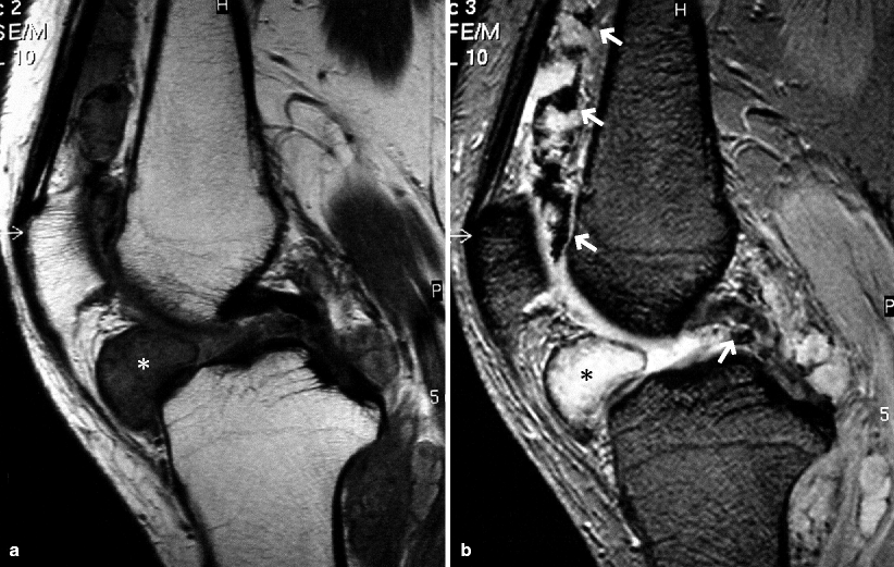
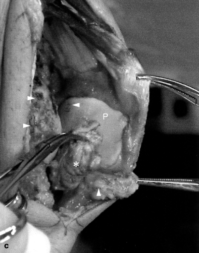
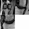
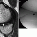
Fig. 11.1
Pigmented villonodular synovitis, localized type. A man in his 50s. (a) PDWI, (b) T2*WI, and (c) intraoperative photo. There is a mass lesion eroding into the Hoffa’s fat pad (*). Within the joint space, there is synovial proliferation with hemosiderin deposition showing hypointensity (arrows). The mass lesion had a yellowish color and proliferated synovium contains black spots representing bleeding (arrowheads)
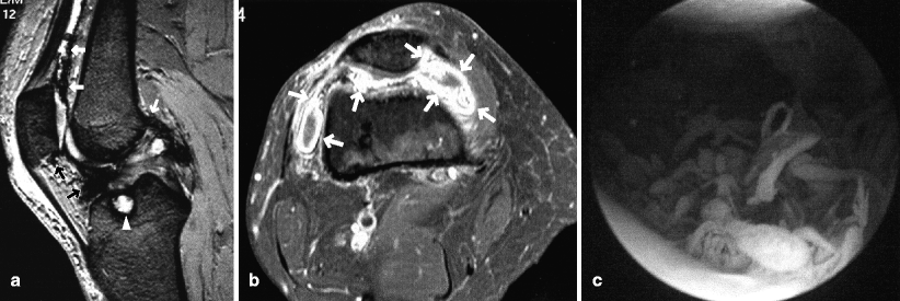
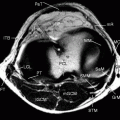
Fig. 11.2
Pigmented villonodular synovitis, diffuse type. A woman in her 50s. (a) T2*WI, (b) axial FS postcontrast T1WI, and (c) arthroscopic image. Within the joint space including the posterior joint capsule, there is diffuse synovial proliferation which shows hypointensity on T2*WI (arrows). There is a cystic change in the tibial plateau due to erosion of the synovium (arrowhead). Proliferated synovium shows strong enhancement after gadolinium injection. Arthroscopically, proliferated synovium has a villous appearance
Reference
Narváez JA, Narváez J, Aguilera C, et al. MR imaging of synovial tumors and tumor-like lesions. Eur Radiol. 2001;11:2549–60.
11.2 Giant Cell Tumor of Tendon Sheath
This is histologically identical to PVS described in the previous section.
It mostly arises in the tendon sheath. In the knee joint, it commonly arises adjacent to the joint capsule.
In old days it was called “xanthoma.”
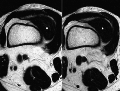
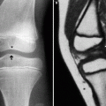
Fig. 11.3
Giant cell tumor of tendon sheath. A woman in her 50s. Axial T2WI (left) and T1WI (right). There is a mass lesion (*) which generally shows hypointensity with some faint hyperintense component on T2WI adjacent to the suprapatellar bursa. Histologically, it is proliferated synovium, which is identical to that found in PVS
11.3 Synovial Osteochondromatosis
It is a benign condition of unknown etiology characterized by synovial nodular proliferation containing cartilaginous and osseous components.
Common in young to middle-aged persons.
Knee, hip, and elbow joints are most commonly affected.
Chondroma may grow into the joint space, and fragments may break off from the synovial surface into the joint, where they may enlarge, calcify, or ossify. Appearance of such loose fragments resembles that of rice bodies seen in rheumatoid arthritis.
Intra-articular proliferation of osteochondroma and multiple loose fragments causes widening of the joint space and overall swelling of the joint.
Erosion of bone cortex may be seen.
Usually monoarticular.
If loose fragments ossify or calcify, they can be visualized on radiograph. However, this is only the case in 30–40% of cases.
Clinically, patients initially have dull pain and swelling due to joint effusion. When loose fragments drop into the joint space, range of movement of the affected joint becomes limited and may eventually lead to osteoarthritis. Synovial proliferation is said to cease spontaneously.
Treatment includes excision of proliferated synovium and removal of loose fragments under arthroscopy. If complete removal is not possible arthroscopically, open surgery is required. If actively proliferating synovium is not completely removed, postoperative recurrence may occur, but prognosis is good in general.
Key points for MRI interpretation
Intra-articular soft tissue due to proliferation of osteochondroma is seen.
Large osteochondroma may show heterogeneous signal intensities and may include signal void corresponding to ossified component.
Cartilaginous component shows signal intensity similar to that of articular cartilage.
If sufficient contrast between the cartilaginous component and joint effusion cannot be obtained, MTC technique should be applied.
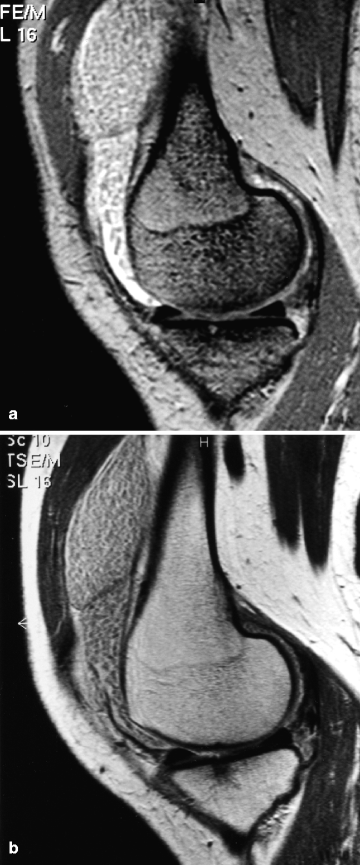
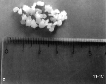
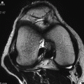
Fig. 11.4
Synovial chondromatosis. A woman in her 30s. T2*WI with MTC, (b) sagittal postcontrast T1WI taken 2 h after gadolinium injection, and (c) excised surgical specimen. Within the joint capsule, there are numerous micronodules that show signal intensity similar to that of articular cartilage (a). Contrast between the nodules and joint fluid can be obtained by using either MTC technique (a) or contrast-enhanced MRI (b) in which gadolinium diffuses into the joint fluid in a late phase. Proliferating synovium and loose fragments were excised arthroscopically. Numerous rice body-like micronodules (a few millimeters in size) were removed from the joint space. In this case, radiograph and CT did not reveal any ossified components, and diagnosis of synovial chondromatosis was made
Reference
Narváez JA, Narváez J, Ortega R, De Lama E, Roca Y, Vidal N. Hypointense synovial lesions on T2-weighted images: differential diagnosis with pathologic correlation. AJR. 2003;181:761–9.
11.4 Synovial Hemangioma
Synovial hemangioma arising within the knee joint is rare.
Histologically, it is commonly a cavernous hemangioma, or a mixture of cavernous and capillary hemangioma.
Common in young persons.
Repetitive intra-articular hemorrhage cause knee swelling, pain, and restricted range of motion.
Radiograph may reveal a phlebolith.
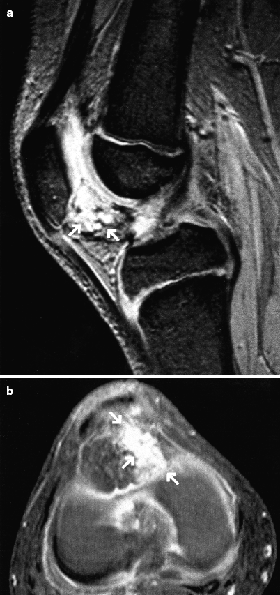
Fig. 11.5
Synovial hemangioma. An 8-year-old girl with history of recurrent hemarthrosis. (a) T2*WI and (b) axial gadolinium-enhanced FS T1WI. There is a multilocular mass showing hyperintensity on T2*WI eroding into Hoffa’s fat pad (arrows, a). The lesion shows strong enhancement after gadolinium injection (arrows, b)
Key points for MRI interpretation
Related posts:
Stay updated, free articles. Join our Telegram channel

Full access? Get Clinical Tree


