George M. Bridgeforth, Joan Williams, and Charles Carroll IV
A 83-year-old woman fell in her kitchen and tried to break her fall with her right hand.
CLINICAL POINTS
- A triangular fibrocartilage complex (TFCC) tear may present with pain and soreness along the distal ulna.
- Pain with torquing (i.e., opening jars or turning keys) is common.
- Patients may present with clicking with wrist movement.
- Triangular fibrocartilage tears may present with pain and soreness along the distal ulna.
- Carpal tunnel symptoms often occur with distal radius fractures.
Clinical Presentation
Distal radial fractures account for approx-imately 17.5% of all fractures. The most common mechanism of injury is a fall onto outstretched hand usually with the wrist in dorsiflexion. In younger individuals, fractures may result from higher energy traumas.
Other injuries from falls unto the outstretched hand are more commonly missed. Scaphoid fractures occur almost as commonly as distal radial fractures (see Chapter 39, “Scapholunate Dissociation”). Clinically, distal radial fractures are characterized by pain, swelling, and tenderness of the anatomic snuffbox and scaphoid region just distal to the radius.
A dislocation of the carpal ligaments may occur following a fall, and the clinician may easily miss this. Usual characteristics include moderate-to-marked pain and soreness between the scaphoid and the lunate at the center of the proximal wrist. Patients may complain of clicking. A Watson shift test may reveal a subluxating scaphoid. The test is performed by stabilizing the patient’s wrist with one hand and placing the patient’s wrist in ulnar deviation. The examiner stabilizes the wrist with the opposite hand and places his or her thumb over the volar scaphoid. He or she then places the patient’s wrist in extension with ulnar deviation and then applies pressure as the wrist is flexed and radially deviated repeatedly. A scapholunate dislocation with a rotating subluxating scaphoid produces a noticeable click. However, other injuries such as a perilunate dislocation or a TFCC tear (distal ulna) may cause clicking of the wrist as well.
Radial and ulnar styloid fractures may be characterized by focal tenderness and pain with limited range of motion. Examiners can easily miss them on radiographs. In fact, the presence of an ulnar fracture should prompt reexamination of the radiographs for an associated radial fracture. Patients with persistent pain and soreness over the wrist who do not have a scaphoid or distal radius fracture may develop de Quervain’s tendonitis. In the absence of a fracture, it is a tendonitis of the extensor and abductor tendons of the thumb. A positive Finkelstein’s test (pain over the anatomic snuffbox with ulnar deviation of the wrist) is characteristic. Treatment includes analgesics, spica splints, cold packs, physical therapy, and steroid injections.
PATIENT ASSESSMENT 
- Pain, swelling, and tenderness of the wrist
- Pain-limited range of motion and grip strength
Finally, falls on the ulnar wrist with the hand pronated may result in damage to the ulnar nerve as it passes through Guyon’s canal. Although these injuries are not common, they can be disabling. It is important to check for tenderness over the hypothenar eminence as well as for ulnar damage (weakness of the interosseous muscles, and flexor digitorum profundus of the fourth and fifth fingers associated with numbness of the hypothenar eminence and the ulnar border of the ring finger and the little finger).
NOT TO BE MISSED 
- Scaphoid and other carpal fractures
- Perilunate dislocation
- Hypothenar pain
- Ulnar nerve palsy from damage to Guyon’s canal
When performing the physical examination, the physician should:
- palpate the distal radius, ulna, scaphoid, and interosseous region for focal tenderness and bony displacement.
- evaluate the ipsilateral elbow and shoulder for associated injuries.
- perform careful neurovascular assessment, paying particular attention to the median nerve. Carpal tunnel symptoms are common with distal radius fractures due to traction during hyperextension or increased compartment pressure secondary to swelling.
- check for clicking (subluxated carpal bone or a TFCC tear).
- check neurovascular status (cold cyanotic limb with diminished or absent pulses)
- check for ganglion cysts.
- check the hypothenar eminence for ulnar nerve injuries (hypothenar numbness with numbness and weakness of the fourth and fifth fingers). Evaluate for hypothenar pain; look for fractures in the hook of the hamate and hamate, as well as pisiform and triquetral fractures.
COLLES AND SMITH FRACTURES
A Colles fracture is the most common form of distal radius fracture, accounting for 90% of distal radius fractures. Most of these fractures can be recognized by the marked swelling, pain, and tenderness of the wrist. In addition, the dorsally displaced distal fragment causes a characteristic deformity of the distal forearm, known as the “dinner fork” deformity. Wrist range of motion is pain limited.
A Smith fracture, sometimes known as a reverse Colles fracture, is another type of fracture of the distal radius. This type of injury is typically unstable. It is characterized by volar (ventral) displacement of the distal radius. (In a Colles fracture, the displacement is dorsal.) The volar displacement may be caused by a fall on a flexed wrist.
BARTON FRACTURES
Barton fractures, which are uncommon, are fractures involving the volar or palmar rim (articulation) of the distal radius. They occur as the result of the carpal bones shearing against the volar or dorsal lip of the radial articular surface. Although Barton fractures may appear stable, they can be associated with torn carpal ligaments and carpal instability. In addition, radial styloid fractures are often seen in association with these fractures. (Radial styloid fractures are commonly linked with intercarpal ligamentous injuries such as scapholunate dissociation or perilunate dislocation.)
Radiographic Evaluation
It is necessary to obtain posteroanterior (PA) and lateral views (Fig. 37.1) of the wrist, along with oblique views for further fracture definition, if needed. Optional tests include

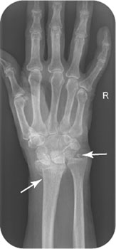
FIGURE 37.1 Lateral radiograph of the right wrist of the patient in the introductory case, demonstrating acute fracture of the right distal radius and ulnar styloid.
Related posts:


Stay updated, free articles. Join our Telegram channel

Full access? Get Clinical Tree


