Nausea and vomiting, weight loss, anemia, upper GI bleed
Periampullary tumors may present with jaundice
• Rare: Represents < 1% of all gastrointestinal neoplasms
DIAGNOSTIC CHECKLIST
• Most duodenal carcinomas cause focal stenoses or obstruction
• A large mass with cavitation is more likely to be lymphoma or GIST
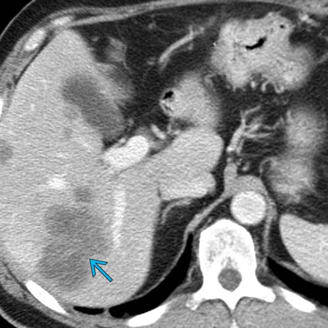
(Left) Axial CECT in a 60-year-old man with weight loss and early satiety shows obvious liver metastases .
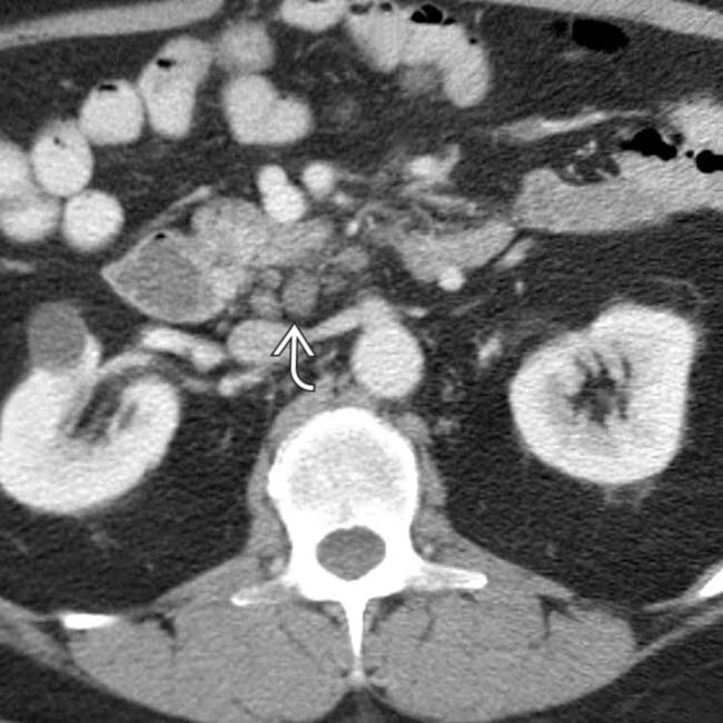
(Right) Axial CECT in the same patient also shows paraduodenal lymph node metastases .
(Left) Axial CECT in the same patient shows the relatively subtle mass that narrows the 3rd portion of the duodenum . There is also a subtle extension of tumor along the superior mesenteric vessels .
(Right) Film from an upper GI series in the same patient shows the duodenal carcinoma more clearly. Note the “shoulder” or abrupt transition to tumor at its proximal extent. The lumen of the more proximal duodenum is dilated.
TERMINOLOGY
Abbreviations
• Duodenal carcinoma (CA)
Synonyms
• Duodenal adenocarcinoma
Definitions
• Primary malignant neoplasm arising in duodenal mucosa
IMAGING
General Features
• Best diagnostic clue
Irregular intraluminal mass or “apple core” lesion at or distal to ampulla of Vater
• Location
15% in 1st portion of duodenum
40% in 2nd portion of duodenum
45% in distal duodenum
• Size
Usually < 8 cm
• Morphology
Polypoid, ulcerated, or annular constricting mass
Intraluminal mass with numerous frond-like projections for carcinomas arising in villous tumors
Radiographic Findings
• Radiography
Proximal obstruction pattern if lumen severely narrowed
Fluoroscopic Findings
• May have various appearances
Ulcerated mass
Polypoid mass
Annular constricting “apple core” lesion
“Soap bubble” reticulated pattern for villous tumors
CT Findings
• CECT
Discrete mass or irregular thickening of duodenal wall
Concentric narrowing of duodenal lumen
Polypoid intraluminal mass
Local lymphadenopathy
Infiltration of adjacent fat
Biliary ± pancreatic duct dilatation
– With periampullary tumors
Liver ± peritoneal metastases
MR Findings
• MRCP
May see pancreatic or biliary ductal dilatation with periampullary duodenal carcinomas
Ultrasonographic Findings
• Grayscale ultrasound
Hypoechoic mass in duodenum with echogenic center: Pseudokidney sign
• Color Doppler
May see invasion of adjacent vascular structures
Imaging Recommendations
• Best imaging tool
Thin-section CECT with water for luminal distention and dual-phase arterial and venous imaging
• Protocol advice
Multiplanar MIP and volume-rendered CT images
DIFFERENTIAL DIAGNOSIS
Neoplasms
• Ampullary and periampullary adenocarcinomas
Pancreatic ductal carcinoma
– Hypodense mass centered in pancreas with ductal obstruction
Ampullary carcinoma
Cholangiocarcinoma
– Biliary obstruction with small mass
Only gold members can continue reading. Log In or Register to continue


 .
.
 .
.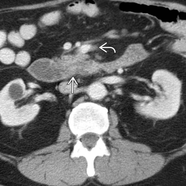
 . There is also a subtle extension of tumor along the superior mesenteric vessels
  .
.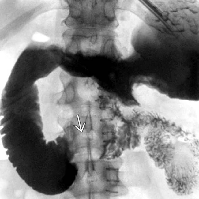
 more clearly. Note the “shoulder” or abrupt transition to tumor at its proximal extent. The lumen of the more proximal duodenum is dilated.


