George M. Bridgeforth, David S. Wellman, and Charles Carroll IV
A 20-year-old man was wrestling And heard his arm “pop” while wrestling. He presents with severe elbow pain and cannot bend his arm.
CLINICAL POINTS
- The majority of elbow dislocations involve posterior displacement.
- Some deformity may be present.
- It is important to examine the middle and distal forearm for an associated fracture.
A thorough physical examination is essential. The physician should:
- look for marked pain, swelling, tenderness, and deformity.
- check for limited range of motion with crepitus.
- check for neurovascular impairment (i.e., cold limb, with diminished or absent radial, ulnar, and brachial pulses; dusky hue).
- check skin integrity.
- evaluate for ulnar nerve damage: weakness of the ulnar wrist flexors and interosseus muscles, as well as sensory impairment of the hypothenar eminence and the fourth (ulnar half) and the fifth finger.
- evaluate for median nerve damage: weakness of the radial wrist flexors and thumb interphalangeal joint flexion with a sensory impairment of the thenar eminence and the volar first, second, third, and radial half of the fourth fingers.
Clinical Presentation
The elbow is a relatively stable hinge joint, and dislocation of this joint requires considerable force. Dislocation of the elbow is second in frequency to that of the shoulder. Athletic injuries account for up to 50% of elbow dislocations. Posterior dislocations are most common (90%) and may result from a fall onto an outstretched hand with a combination of axial, rotational, and varus (or valgus) force. For example, a person who is ice skating may fall backward and extend an arm to break his or her fall. Anterior dislocations occur much less frequently as a result of direct trauma to the flexed elbow. Associated fractures often occur with elbow dislocations.
An elbow dislocation is not difficult to diagnose; the elbow deformity is readily evident and is associated with a marked pain, swelling, and tenderness of the elbow. Impaired range of motion also occurs.
A plain radiographic workup should follow the initial physical examination (see section, “Radiographic Evaluation”).
Finally, the clinician should evaluate the patient for evidence of the “terrible triad.” This consists of an elbow injury with radiographic evidence of a radial head fracture and a coronoid fracture. The terrible triad occurs in approximately 10% of elbow dislocations and is more common with posterior dislocations. If the physician misses a terrible triad injury, the fracture of the coronoid may result in recurrent elbow subluxations due to hinge instability.
Radiographic Evaluation
A complete elbow series consists of anteroposterior, lateral, and oblique radiographs of the elbow, and these diagnose most dislocations and subluxations. The clinician should evaluate each film closely as a subluxation can be subtle. A view specifically centered on the radial head and capitellum can be obtained if there is concern about radial head or capitellum fracture/dislocations.
Dislocations can be simple or represent components of fracture dislocations with complex associated injuries. There may be fractures, dislocations, subluxations, and ligament injuries, all occurring in the same setting. A computed tomography (CT) scan or magnetic resonance imaging scan may be ordered to evaluate these injuries further and to assist with preoperative planning; however, the dislocated joint should be reduced first.

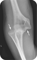
FIGURE 34.1 Lateral radiograph of the patient in the introductory case, showing a left elbow dislocation.
Related posts:


Stay updated, free articles. Join our Telegram channel

Full access? Get Clinical Tree


