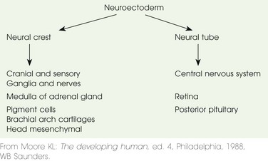
Chapter 24 The human brain undergoes four phases of development: (1) dorsal induction (primary and secondary neurulation), (2) ventral induction (patterning of the forebrain), (3) neuronal proliferation and migration, and (4) myelination. During the third week of embryogenesis, initiation of the central nervous system evolves with the development of the notochordal process. This derivative of ectoderm grows rapidly in length so that by 20 days it is converted from a hollow tube to a solid rod—the notochord (Fig. 24-1; e-Table 24-1). The notochord works with the axial mesoderm to induce the neural plate. The neuroepithelium of the neural plate begins the formation of the brain and spinal cord. It appears initially at the cranial end of the embryo and differentiates craniocaudally. At the beginning of the fourth week, the neural plate is composed of a broad cranial portion and a narrow caudal portion—the fetal brain and spinal cord. Figure 24-1 Further development of the notochord by transformation of the notochordal process. e-Table 24-1 Schematic Chronology of the Major Events During Human Neocortical Development From Gressens P, Helppi PS. Normal and abnormal brain development. In Martin R, Fanaroff A, Walsh M, eds. Neonatal-perinatal medicine. 8th ed. Elsevier Mosby; 2005. The process of neurulation or formation of the neural tube occurs when the lateral edges of the neural plate elongate to become neural folds and join together to form the neural tube (Fig. 24-2).1 The process starts at the craniocervical region and proceeds superiorly and inferiorly. The space within the neural tube, the neural canal, initially is open at the cranial (or rostral) and caudal ends (cranial and caudal neuropores) and communicates with the amniotic cavity. The neuropores gradually decrease in size and close between the twenty-fourth and twenty-sixth day, apparently at multiple sites and not necessarily in a craniocaudal fashion.1 The lowest portion of the spinal cord, the inferior sacral and coccygeal levels, are formed by a different process called secondary neurulation (see Chapters 40 and 43). Pluripotent tissue within the caudal eminence forms a solid neural cord; this neural cord forms a lumen that fuses with the neural tube (e-Fig. 24-3). This secondary process is not completed until the eighth week after fertilization of the ovum.1 Figure 24-2 Drawings of embryos of 19 to 21 days, illustrating development of the somites and intraembryonic coetom. e-Figure 24-3 Formation of the neural tube inferior to the second sacral level by secondary neurulation. Some neural crest cells (the population of neural cells arising at the lateral edge of the neural plate during neural tube formation) detach during neurulation and form many different tissues (e.g., melanocytes and chromaffin cells of the adrenal medulla), including the major components of the peripheral and autonomic nervous system (e-Table 24-2).1 The prechordal plate cephalic to the neural tube and notochord induces this stage of development. The three major divisions of the brain—the prosencephalon or forebrain, mesencephalon or midbrain, and rhombencephalon or hind brain—are more clearly differentiated during the rostral expansion of the neural tube (the formation of the primary brain vesicles).1,2 During the fourth and fifth weeks, a series of brain foldings occur (brain flexures—midbrain, cervical, and later pontine) so that by the end of the fifth week, five secondary brain vesicles are present (Table 24-3). With further development, the prosencephalon becomes subdivided into the telecephalon and the diencephalon. The rhombencephalon subdivides into the metencephalon and myelencephalon. The mesencephalon does not divide. Within each of these five vesicles, the neural canal expands into a primary ventricle. The central canal of the spinal cord is continuous with the brain ventricles. The fate of these structures is shown in Table 24-3. The cranial nerve nuclei appear in the brainstem during the fifth week (see the next section).1
Embryology and Brain Development
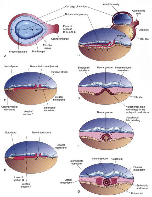
A, Dorsal view of the embryonic disc (about 18 days), exposed by removing the amnion. B, Three-dimensional median section of the embryo. C and E, Similar sections of slightly older embryos. D, F, and G, Transverse sections of the trilaminar embryonic disc shown in C and E. (From Moore KL, Persaud TVN. Before we are born. 5th ed. Philadelphia: WB Saunders; 1998.)
Event
Time Event Occurs
Neuroectoderm induction
Third GW
Neurulation
Third to end of fourth GW
Proencephalic and hemispheric formation
Fifth to tenth GW
Neuronal proliferation
Tenth to twentieth GW
Neuronal migration
Twelfth to twenty-fourth GW
Programmed neuronal cell death
Twenty-eighth to forty-first GW
Neurogenesis
Fifteenth to twentieth GW to ? postnatal months or years
Synaptogenesis
Twentieth GW to puberty
Gliogenesis
Twenty to twenty-fourth GW to ? postnatal years
Myelination
Twenty-sixth to twenty-eighth GW to 2 to 3 postnatal years
Angiogenesis
Fifth to tenth GW to ? postnatal years
Dorsal Induction
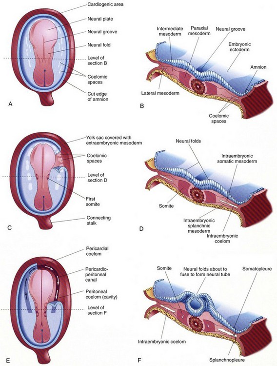
A, C, and E, Dorsal view of the embryo, exposed by removal of the amnion. B, D, and F, Transverse sections through the embryonic disc at the levels showing. A, Presomite embryo of about 18 days. C, An embryo of about 20 days, showing the first pair of somites. A portion of the somatopleure on the right has been removed to show the isolated coelomic spaces in the lateral mesoderm. E, A three-somite embryo (about 21 days) showing the horseshoe-shaped intraembryonic coelom, exposed on the right by removal of a portion of the somatopleure. (From Moore KL, Persaud TVN. Before we are born. 5th ed. Philadelphia: WB Saunders; 1998.)
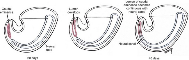
Mesoderm invading this region during gastrulation condenses into a solid rod called the caudal eminence, which later develops a lumen. At the end of the sixth week, this structure fuses with the neural tube. (From Larsen WJ. Human embryology. 2nd ed. New York: Churchill Livingstone; 1997.)
Ventral Induction
![]()
Stay updated, free articles. Join our Telegram channel

Full access? Get Clinical Tree


Radiology Key
Fastest Radiology Insight Engine