12 Estimating Fetal Gestational Age The gestational age assignment to a pregnancy is needed subsequent to evaluation to assess the fetal anatomy and growth, interpret the various screening tests, and predict the expected delivery date. There are various ways of calculating the fetal gestational age, including menstrual history, clinical examination, and ultrasound.1–3 Of these three, ultrasound is superior for dating a pregnancy.3 The menstrual age is calculated from the first day of the last menstrual period. The conceptual age is calculated from ovulation. The gestational age is calculated from the theoretical time of ovulation, plus 2 weeks. In addition to estimating fetal gestational age, fetal biometry is used to detect problems related to growth disturbances, such as intrauterine growth retardation, macrosomia, and microcephaly. The other advantages of fetal biometry are the early diagnosis of malformations (e.g., the measurement of long bones in skeletal dysplasias)4 and detection of patients at risk for aneuploidy (fetal nuchal translucency).5 Calculations based on fetal measurements are also used to determine growth and weight. Ultimately, assessment of fetal biometry is important for decreasing the perinatal morbidity and mortality. The assessment of gestational age and fetal growth is based on different parameters, which are described in this chapter. In all the tables outlined, the gestational age is expressed in weeks and days from the (theoretical) onset of the last menstrual period (conception date minus 14 days).2 There are many well-established charts that have been in use for a long time; however, marked differences between populations sometimes force researchers to build new nomograms for the different races.6 This parameter is useful in the early stages of pregnancy when the fetal pole or the embryo is still not identifiable on ultrasound. This can be as early as 5 weeks by transvaginal examination and 6 weeks by transabdominal exam. In the setting of an ectopic gestation, a pseudogestational sac may mimic a true sac. Therefore, in the absence of a definite fetal pole or yolk sac, caution should be taken in labeling a true sac as such. A normal gestational sac can be confidently diagnosed when its hyperechoic rim is thick (> 2 mm) and its shape is round or oval without unusual angulation. The mean gestational sac diameter is measured inside its hyperechoic rim. If the sac is round, only one measurement is needed. More commonly, when it is oval, three orthogonal (perpendicular) measurements of the sac are taken and averaged. Gestational age is estimated based on the mean sac diameter (Table 12–1). According to one study by Muller et al, three-dimensional volumetric scans are more accurate than two-dimensional scans in obtaining the gestational sac volume, but have shown to be of no prognostic significance for gestational outcome.7
Perameters Used in Fetal Biometry to Determine Gestational Age
Mean Gestational Sac Diameter
Mean Sac Diameter (mm) | Gestation in Weeks (Mean) |
2 | 5.0 |
3 | 5.1 |
4 | 5.2 |
5 | 5.4 |
6 | 5.5 |
7 | 5.6 |
8 | 5.7 |
9 | 5.9 |
10 | 6.0 |
11 | 6.1 |
12 | 6.2 |
13 | 6.4 |
14 | 6.5 |
15 | 6.6 |
16 | 6.7 |
17 | 6.9 |
18 | 7.0 |
19 | 7.1 |
20 | 7.3 |
21 | 7.4 |
22 | 7.5 |
23 | 7.6 |
24 | 7.8 |
25 | 7.9 |
26 | 8.0 |
27 | 8.1 |
28 | 8.3 |
29 | 8.4 |
30 | 8.5 |
The mean gestational age was calculated from a regression equation. Reported range: ± 0.1 week at 2 SD.
From Alfred B. Kurtz. Estimating gestational age. In: Bluth EI, Arger PH, Benson CB, Ralls PW, and Siegel MJ, eds. Ultrasound: A Practical Approach to Clinical Problems. New York: Thieme Medical Publishers; 2000. Reproduced with permission.
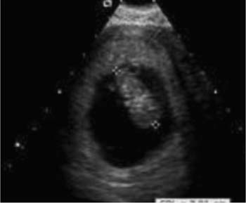
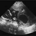
Figure 12–1 Scan demonstrating the crown–rump length measurement.
Crown–Rump Length
The crown–rump length (CRL) is the longest demonstrable length of the embryo, excluding the limbs and yolk sac.8The CRL is the most accurate indicator of the gestational age throughout the first trimester because, early in the pregnancy, the CRL has a rapid linear growth acceleration curve, and, at this stage, the growth of the embryo is minimally affected by various pathologies.
The CRL is measured from the outer edge of the cephalic pole to the outer edge of the fetal rump. One must be careful not to include the yolk sac in the measurement9 (Fig. 12–1).
The CRL can be used to assess the gestational age between 6 and 14 weeks (Table 12–2). Its best accuracy is from 6 to 10 weeks, during which time it has an error of ± 3 to 4 days (95% confidence interval). However, as the gestation advances, there are methodological issues because the fetus may flex and extend, making reproducible linear measurements difficult. The error increases to ± 5 days between 10 and 14 weeks of gestation.
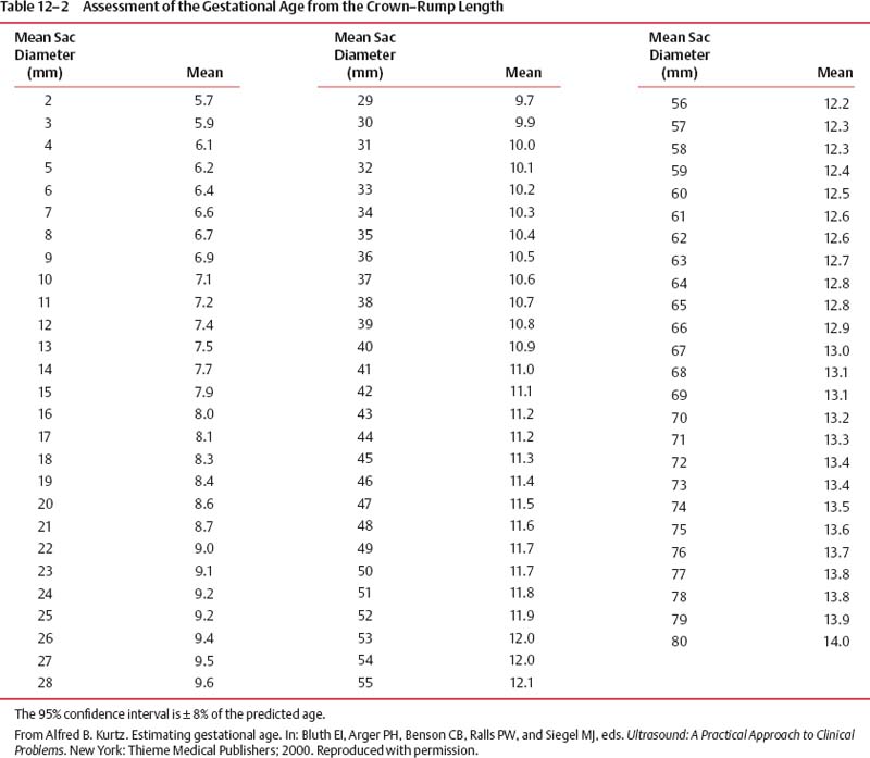
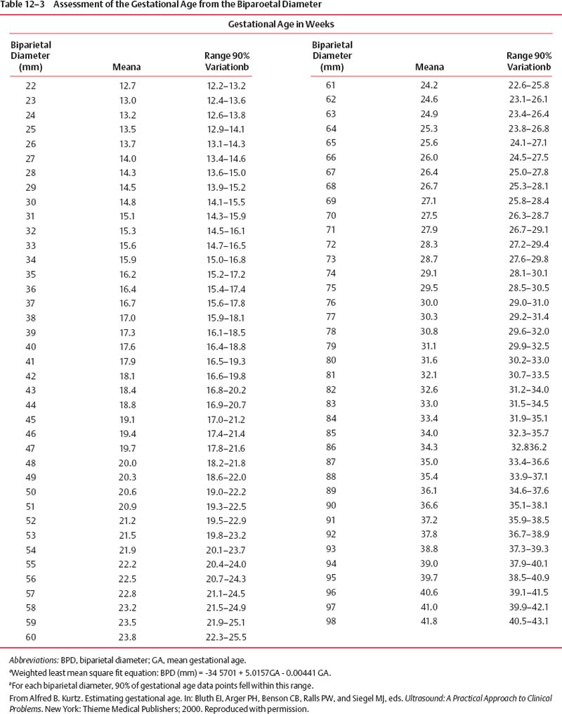
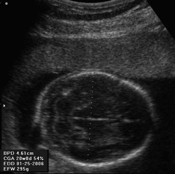
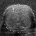
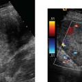
Figure 12–2 Scan demonstrating the biparietal diameter measurement, which is measured from the outer table of the skull to the inner table at the level of the thalami.
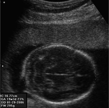
Figure 12–3 Scan demonstrating the measurement of the head circumference.
The CRL parameter in the first trimester is as accurate as or more accurate than any second-trimester fetal measurement for estimating gestational age.10 There are no well-established measurement tables of fetal parts for gestational ages below 12 weeks.
Biparietal Diameter
The biparietal diameter (BPD) is one of the first parameters that was used to estimate gestational age and has been extensively researched in the literature. BPD is the most widely accepted parameter in estimating fetal gestational age11,2 (Table 12–3). It is measured in a transverse plane at the level of the thalami from the outer table of the proximal skull to the inner table of the distal skull, corresponding to the “leading edge to leading edge” measurement (Fig. 12–2).
Measurements rostral to the thalami (i.e., below the level of the cerebral peduncles) may result in underestimation of the BPD and consequently the fetal gestational age. For accurate serial assessment, the biparietal diameter should be measured using the same landmarks (i.e., level of the thalami). If the BPD is measured at different levels, erroneous readings can result.
The biparietal diameter is most accurate between 12 and 28 weeks of gestation. After 28 weeks of gestation, head shape, intrauterine crowding, and individual variations affect the size of the head; hence, BPD must be used with caution.2
Difficulties Accurately Measuring the Biparietal Perimeter
Occasionally, the fetal head may be dolichocephalic (flattened and elongated) or brachycephalic (broadened and shortened), resulting in inaccurate estimation of gestational age based on BPD measurements.13
Related posts:
Stay updated, free articles. Join our Telegram channel

Full access? Get Clinical Tree


