(1)
Department of Radiology, Saitama Medical University, Moroyama, Saitama, Japan
Abstract
Tibial plateau fracture (fracture of proximal end of tibia) is one of the most common traumatic fracture of the knee joint.
8.1 Tibial Plateau Fracture
Tibial plateau fracture (fracture of proximal end of tibia) is one of the most common traumatic fracture of the knee joint.
It is commonly seen in osteoporotic elderly patients, but also frequently seen as traumatic or sports injuries.
Tibial fracture results following collision of the distal femur and the tibial plateau due to external forces.
Valgus stress causes fracture of the lateral condyle and commonly accompanied by tear of MCL and cruciate ligaments. Hemarthrosis may also result.
In clinical practice, Hohl’s classification is commonly used (Fig. 8.1).
Hohl’s undisplaced type and other types with little displacement of the fractured bone (Fig. 8.2) are treated conservatively.
If the lesion includes vertical fracture of the medial or lateral condyle, it requires manipulation and fixation. If there is a depression fracture involving the joint surface, surgical fixation is required (Fig. 8.3).
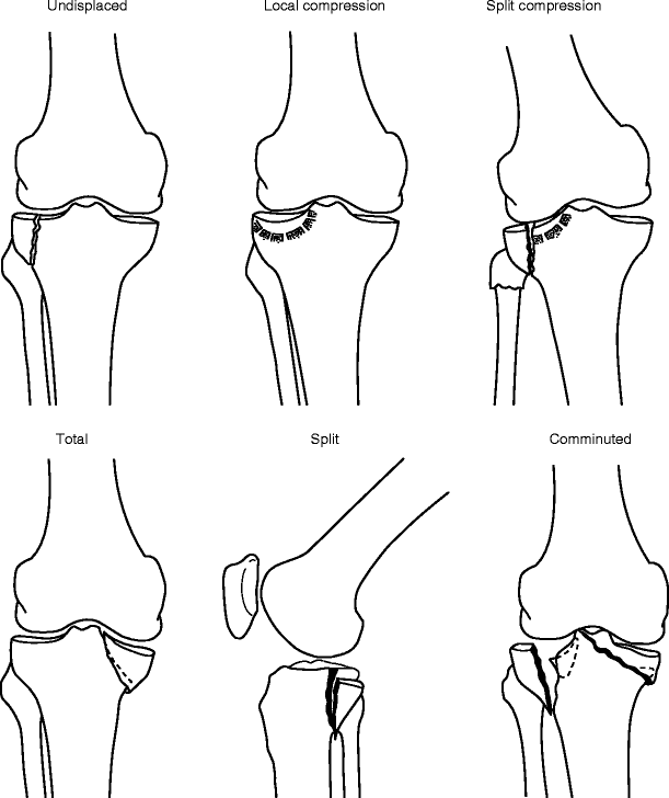
Fig. 8.1
Hohl’s classification of the tibial plateau fracture (Images adapted from Hohl’s publication)
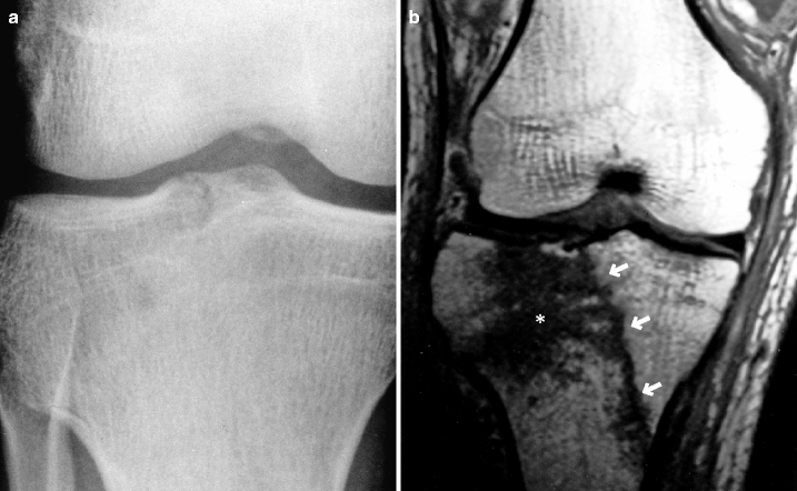
Fig. 8.2
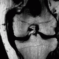
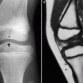
Tibial plateau fracture, undisplaced type. A man in his 20s. (a) Anteroposterior radiograph and (b) coronal T1WI. This is an example of a tibial plateau fracture, undisplaced type, according to the Hohl’s classification. It is not clearly visualized on radiograph, but there is a clear linear hypointensity running obliquely from the intercondylar eminence (arrows) on MRI. Bone bruise is noted (hypointensity on T1WI (*, b) and hyperintensity on T2WI (not shown))
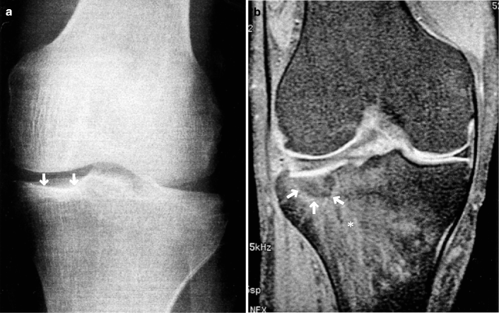
Fig. 8.3
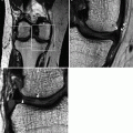
Tibial plateau fracture, local compression type. A man in his 50s. (a) Anteroposterior radiograph and (b) coronal FS T1WI. Radiograph depicts depression of the joint surface (arrows), while MRI demonstrates clearly a fracture line (arrows) together with extensive bone bruise (*, b)
8.1.1 Reference
Hohl M. Tibial condylar fractures. J Bone Joint Surg. 1967;49-A:1455–67.
8.1.2 Patella Is the Largest Sesamoid Bone of the Human Body
This can be common sense, but perhaps some people have never thought of it this way. A sesamoid bone is a small circular bone which is embedded within a tendon. I am sure people often think of sesamoids in the hands and feet. Patella is a large sesamoid bone that has hyaline cartilage because it is involved in the patellofemoral joint. Another well-known sesamoid in the knee is the fabella, which is found within the lateral head of the gastrocnemius. A ligament called fabellofibular ligament also exists.
8.2 Patellar Fracture
Patellar fracture can be classified as transverse fracture and comminuted fracture. Transverse fracture is more common with a frequency of more than 50%.
Transverse fracture results from a sudden flexion of the knee. Due to eccentric contraction of the quadriceps femoris muscle, patella is torn into two pieces (superior and inferior fragments). The bone fragments are commonly displaced to a large extent.
Treatment plan will be made depending on the degree of separation of the bone fragments on lateral radiograph taken at knee extension. If the separation is insignificant, conservative treatment will be instituted.
Comminuted fracture of the patella results from a direct external force applied from the anterior direction, such as a fall or a dashboard injury. In this case, bone fragments tend to remain in the original position.
Patellar fracture is an intra-articular fracture and thus commonly accompanied by hemarthrosis (Fig. 8.5).
Patellar fracture in children is rare. If it occurs, it takes the form of patellar sleeve fracture, which is an avulsion fracture of the inferior pole of the patella.
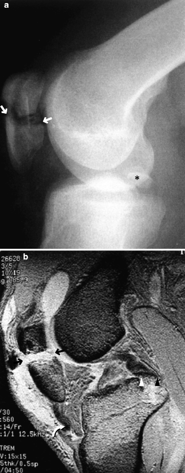
Fig. 8.4
Patellar fracture. A man in his 20s following a traffic accident. (a) Lateral radiograph and (b) T2*WI. The patella is split into two large fragments (white arrows in a and black arrows in b mark the space between two fragments). MRI shows kinking of the patellar tendon (curved arrow) and the avulsion fracture of the PCL (arrowheads, b) * avulsed bone fragment (a)
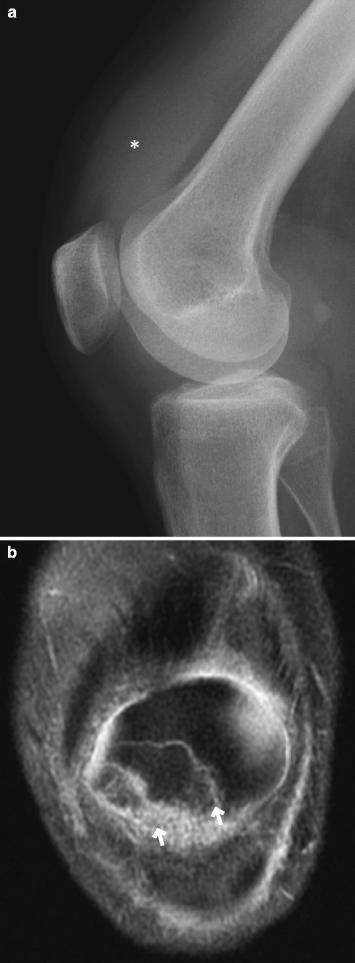
Fig. 8.5
Patellar fracture. A woman in her 50s who had a fall 2 days prior and had hemarthrosis at presentation. (a) Lateral radiograph and (b) coronal FS PDWI. Radiograph shows joint effusion in the suprapatellar bursa (*, a). MRI shows two fracture lines in the patella (arrows, b)
8.3 Patellar Dislocation
Patella may be dislocated from the patellofemoral groove, in which it normally rests. If the joint surface is partially in contact, it is called subluxation.
Depending on the direction of the dislocation, it can be classified as lateral, medial, and horizontal dislocation. The horizontal dislocation is accompanied by torn quadriceps femoris or patellar tendon. Lateral dislocation is most commonly seen (Fig. 8.8).
Patellar dislocation (subluxation) is mostly repetitive. Singularly occurring traumatic dislocation without a known history of repetitive dislocation is rare.
Risk factors for repetitive patellar dislocation include malalignment of the lower limb (e.g., valgus) in general, malalignment of the patellofemoral joint, patella alta, generalized joint laxity, and other congenital and developmental factors.
Hypermobility of the patella may cause instability and pain without causing dislocation. This is called patellar instability.
Patella can be morphologically classified using Wiberg classification (Fig. 8.6).
Repetitive dislocation is common in young women. With the knee lightly flexed, external rotation of the distal lower limb and strong contraction of the quadriceps femoris cause the lateral dislocation/subluxation of the patella (Fig. 8.7).
Traumatic dislocation (acute dislocation) is commonly accompanied by a tear or injury of the medial retinaculum. Traumatic dislocation may occur in persons who are predisposed to repetitive dislocation due to aforementioned risk factors (Fig. 8.8).
Lateral dislocation can often be naturally reduced, and by the time the patient comes to a hospital, the patient has pain but the patella is positioned normally.
Patella dislocation may coexist with the tangential osteochondral fracture (see later description), which can be detected by MRI.
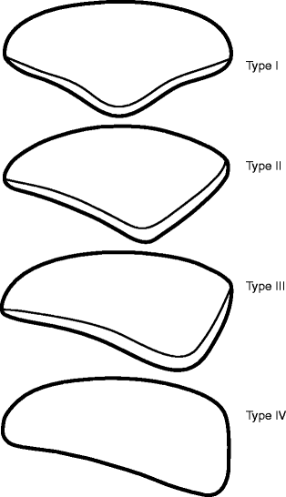
Fig. 8.6
Wiberg classification of the patellar shape. Type I: the facets are concave, symmetrical, and of equal size. Type II: the medial facet is smaller than the lateral facet and flat or only slightly convex. The lateral facet is concave. Type III: the convex medial facet is markedly smaller than the concave lateral facet, and the angle between the medial and lateral facets is nearly 90°. Type IV: The unifacet patella which is also called Jaegerhut shape
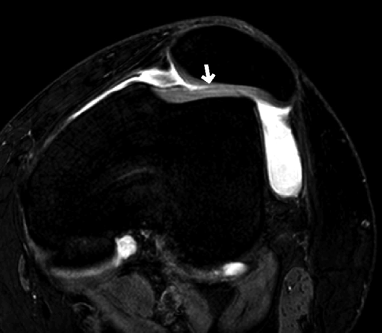
Fig. 8.7
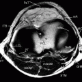
Malalignment of the patellofemoral joint. A woman in her 50s with patellar instability with a history of repetitive patellar subluxation. Axial FS PDWI shows the Wiberg type III patella. Note the thinning of the cartilage of the lateral facet (arrow)
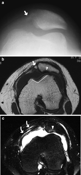
Fig. 8.8
Traumatic lateral patellar dislocation in a patient with known patellar instability and repetitive patellar dislocation. A woman in her late teens. (a) Skyline view radiograph, (b) axial PDWI, and (c) axial FS T2WI. Note the Wiberg type III patella and a shallow patellofemoral groove (bent white line, b). Partial loss of cartilage in the lateral facet and subchondral signal changes are noted (arrowheads, b, c). Medially, there is a small bone fragment (arrow, a, b). Medial retinaculum is torn due to trauma (curved arrow, c)
Reference
Kirsch MD, Fitzerald SW, Friedman H, Rogers LF. Transient lateral patellar dislocation: diagnosis with MR imaging. AJR. 1993;161:109–13.
Patella alta
Patella alta is an abnormally high (proximal) patella in relation to femur and can cause patellofemoral malalignment, increasing the risk of patellar instability including patellar dislocation.
It may also occur as a result of patellar tendon tear or patellar sleeve fracture.
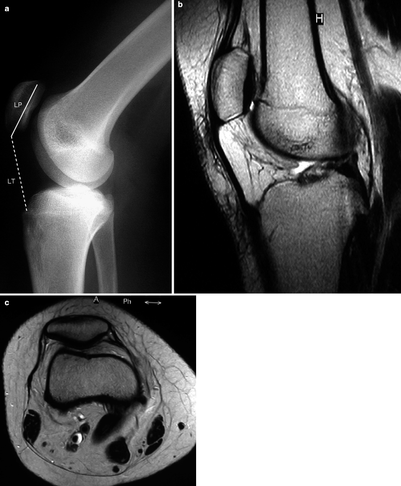
It is defined as Insall-Salvati index ≥1.2. Patella baja is defined as Insall-Salvati index ≤0.8 (Fig. 8.9).

Fig. 8.9
Patella alta. A 14-year-old girl with a history of repetitive patellar subluxation. (a) Lateral radiograph, (b) sagittal, and (c) axial T2WI. Insall-Salvati index (=LT/LP) is greater than 1.2, which is consistent with patella alta. Axial image shows patellofemoral malalignment (Images courtesy of Dr. Ryuji Sashi, Yaesu Clinic, Japan)
Stay updated, free articles. Join our Telegram channel

Full access? Get Clinical Tree


