Avneesh Chhabra, MD, MBA, FACR, Editor
Flavio Duarte Silva, MD, PhD, Editor
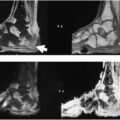
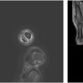
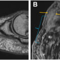
This issue of Radiologic Clinics of North America has the privilege of gathering leading experts in the field of musculoskeletal radiology, attaining comprehensive reviews that enclose both the technical foundations and the diagnostic benefits of high-resolution and 3D extremity and joint imaging. The work highlights the use of advanced techniques for evaluating upper- and lower-extremity intricate anatomy and injury patterns affecting tendons, ligaments, cartilage, nerve, and bone, while also exposing traditional two-dimensional (2D) MR imaging weaknesses that can be overcome by 3D techniques. Several case examples have been shown with comparative findings on 2D and 3D MR imaging so that the reader can gain the needed knowledge of various advantages and pitfalls of such techniques.
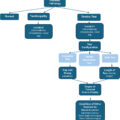
We expect this collection of novel works will serve as an excellent and cutting-edge educational resource and a practical guide for radiologists desiring to explore new frontiers in MR imaging, especially structure-specific reconstructions for superior depiction of anatomy and lesions of fine musculoskeletal structures. Another article on radiology reporting and data systems (RADS) is included that discusses available scoring systems in the domain of musculoskeletal radiology. The readers can use that knowledge for standardized scoring of different musculoskeletal pathologic conditions, including bone and soft tissue tumors and tumorlike lesions, peripheral nerves, and infections. By coupling technological innovation with clinical application, this issue emphasizes the pivotal role of high-resolution and 3D MR imaging in musculoskeletal imaging and its continuing contribution toward optimizing patient care. Enjoy this innovative set of articles in this exciting issue of Radiologic Clinics of North America !
Disclosures
Related posts:
 Detailed Review of MSK Reporting and Data Systems
 3 Dimensional MR Imaging of the Hand and Fingers
 High-Resolution Peroneal Compartment Imaging at Ankle-2d and 3d MR Imaging
 MR Neurography of Lower Extremity Sports-Related Nerve Injuries
 Shoulder Impingement Syndromes: State of the Art Imaging
 Evolving MR Imaging Applications in Posterior Tibial Tendon Dysfunction
Stay updated, free articles. Join our Telegram channel

Full access? Get Clinical Tree


