George M. Bridgeforth, Kevin Bozic, and Jay Patel
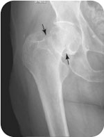
A 97-year-old woman presents with left hip pain after falling.
CLINICAL POINTS
- The elderly are at higher risk for falls. Routinely reviewing medications and arranging for home health visits for geriatric patients are important preventive steps.
- Most (85%) of hip dislocations are posterior (best appreciated on lateral view).
- Hip fractures are divided into intracapsular and extracapsular types.
- A thorough neurovascular examination should always be conducted.
Clinical Presentation
Hips are one of the more common fractured joints; according to Medicare epidemiological studies, hip fractures account for up to 45% of all fractures. The overall cause of hip fractures, as well as hip dislocations, is impact. In individuals younger than 60 years, hip fractures are commonly caused by motor vehicle accidents. In individuals older than 60 years, hip fractures are usually caused by falls. According to the Centers for Disease Control and Prevention, 90% of the hip fractures in adults 65 years of age and older result from falls. There is a higher risk of hip fractions and dislocations in the elderly, especially in Caucasian women with osteoporosis. The mortality rate in the elderly is substantial; 15% to 20% of such patients with hip fractures die within 1 year.
There are two types of hip fractures: intracapsular and extracapsular. Intracapsular hip fractures occur above the intertrochanteric line (a line drawn from the greater to the lesser trochanter on the anteroposterior [AP] view), and extracapsular fractures occur below this point. Intertrochanteric and subtrochanteric fractures occur at or below the intertrochanteric line. Intertrochanteric and subtrochanteric fractures are described according to the number of fractured segments (two to four). The mechanisms of injury involved in hip fractures are
- Subcapital fractures (intracapsular): impaction
- Transcervical fractures (intracapsular): clockwise rotation of the femoral head against a counterclockwise rotation of the femoral neck
- Intertrochanteric fracture (extracapsular): generally from a fall in which there was a direct blow to the hip
- Subtrochanteric fractures (extracapsular): impaction
Approximately 85% to 90% of hip dislocations are posterior, and the remaining are anterior.
The clinical hallmark of a hip fracture is the presence of marked pain, swelling, and tenderness, with an inability to bear weight on the affected side. The clinical examination usually reveals pain-limited range of motion, with marked guarding and apprehension by the patient. It is critical to conduct and document a thorough neurovascular examination that assesses motor weakness, sensory loss, areflexia, and loss of voluntary rectal tone (lower motor neuron injury is generally associated with displaced pelvic fractures). Other causes of the clinical manifestations include hip contusions, avascular necrosis (AVN), degenerative osteoarthritis, and trochanter bursitis.
AVN may be difficult to detect during the initial examination. Radiographic findings may not be apparent for several weeks. In this case, magnetic resonance imaging may help detect an occult fracture or an early AVN. The diagnosis of AVN or an occult fracture should be suspected in any patient who presents with severe hip or groin pain that is not improving. The situation may be complicated by the fact many elderly patients have limited ambulation secondary to degenerative osteoarthritis. The incidence of AVN is greater with intracapsular and displaced hip fractures.
One of the major problems is that severe contusions, hip fractures, dislocations, or AVN may be complicated by bleeding into injured hip. It is important to monitor vital signs carefully for evidence of hemodynamic instability. It is essential to critically review the medications for polypharmacy and limit medications that may cause unwarranted sedation, lightheadedness, and dizziness.
Many elderly patients may be taking multiple medications (especially for high blood pressure); therefore, hypotension with tachycardia is common during the initial presentation. A “stat” complete blood cell count that examines the hemoglobin level and the hematocrit level is an important part of the workup.
Radiographic Evaluation
Radiographs should include AP and lateral (frog leg) views. Hip dislocations are appreciated best on the lateral (frog leg) view. With posterior dislocations, the possibility of a fracture of the posterior acetabular lip caused by the impact from the dislocated femoral head should be checked for very carefully. On the AP view, anterior displacement (10%–15% of the dislocations) may be characterized by a slight medial displacement of the femoral head. It is necessary to look for a subtle effacement of the acetabular space (the cartilaginous cushion that separates the acetabulum from the femoral head).
Hip fractures are divided into intracapsular and extracapsular fractures:
- Intracapsular
- Capital (uncommon)
- Subcapital (common)
- Transcervical (uncommon)
- Capital (uncommon)
- Extracapsular
- Intertrochanteric
- Subtrochanteric
- Trochanteric
- Intertrochanteric
PATIENT ASSESSMENT 
- It is necessary to look for marked pain, swelling, and tenderness. Bruising may or may not be present.
- Pain with limited range of motion may be evident.
- Patients may have pain with limited weight-bearing with an impaired gait.
- Neurovascular status, including rectal tone, should be checked carefully. Associated lower motor neuron injuries to the pelvis may cause loss of bowel and bladder function with impaired voluntary rectal tone, acute motor weakness, sensory impairment, and diminished (or absent reflexes) of the affected extremity.
Related posts:


Stay updated, free articles. Join our Telegram channel

Full access? Get Clinical Tree


