Fig. 43.1
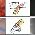
Schema of LVSEA
(a) Sidewall of the lymph vessel is incised with a micro-knife. The incision should be no wider than the outer diameter of the venous stump
(b) Lymph passes (open arrows) from the lymph vessel to the vein and flows proximally
43.1.4 Intraoperative Confirmation of Patency
After completion of the anastomosis, patency can be confirmed by direct visualization through the microscope and by ICG fluorescence lymphography (Fig. 43.2a, b). If the anastomosis is technically well done, lymph will pass from the lymph vessel to the vein, and the vein will become engorged by massage. In cases with good flow of lymph to the vein, the vein can be observed through the skin, representing a good “runoff test” (Fig. 43.2c, d). In my experience, postoperative patency rates seem high in such cases.
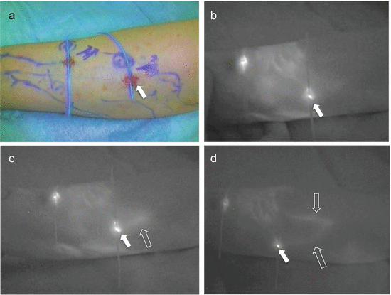
Fig. 43.2
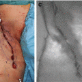
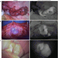
Positive “runoff test” in a 58-year-old woman with chronic lymphedema of the left arm after breast cancer treatment
(a) Two anastomoses were performed in the forearm
(b) ICG fluorescence lymphangiography shows patency of anastomoses in the forearm
(c) Positive “runoff test” of lymph is confirmed by ICG fluorescence lymphangiography (on the right side). Open arrow shows a vein in which ICG is flowing from the lymphatic vessel
(d) The anastomosed vein is observed for longer than in C following manual massage
43.2 Postoperative Evaluations
The primary surgical outcome for lymphatic venous anastomosis is patency of the anastomosis, with volume reduction of the affected limb and improvements in the patient’s activities of daily living (ADL) as secondary and tertiary outcomes, respectively [8]. Patency can be assessed by ICG fluorescence lymphography.
43.2.1 Postoperative Patency
Evaluation by ICG fluorescence lymphography is performed about 6 months after surgery. In my protocol for the treatment of lymphedema, physiotherapy is continued postoperatively for about 6 months, roughly equivalent to the period of preoperative physiotherapy, the content of which is attempted to be reduced thereafter. If the content can be reduced, ADL of the patient increases. Evaluation of patency should therefore be performed at about 6 months after surgery (Fig. 43.3a–d). Through this evaluation, we can clarify the short-term patency. In cases with a patent LVSEA, postoperative ICG fluorescence lymphography shows a Y-shaped pattern of fluorescence (Fig. 43.3c), and branches of the vein also fluoresce (Fig. 43.3d).
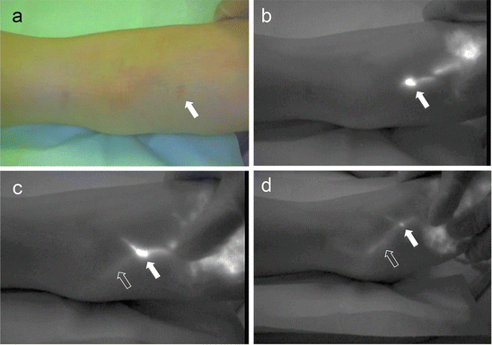
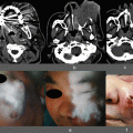
Fig. 43.3
Short-term (6 months after surgery) patency of LVSEA in a 44-year-old woman with right lower-limb lymphedema after treatment for uterine cancer
(a) Closed arrow shows the site of LVSEA in the dorsum of the right foot
(b) ICG fluorescence lymphography shows the lymphatic vessel (linear fluorescence) and anastomosis site (closed arrow)
(c) The anastomosed vein (open arrow) is observed by fluorescence
(d) The anastomosed vein (open arrow) is observed for longer than that in C following massage
As described in this chapter, ICG fluorescence lymphography is limited in its ability to detect the lymphatics and veins selected for anastomosis. If the anastomoses are performed in a relatively superficial layer of subcutaneous tissue, this method can evaluate the patency of the anastomoses. In my study, among 223 anastomoses in the lower limb, only 79 (35 %) could be evaluated by ICG lymphography. The remaining 144 anastomoses could not be evaluated because the subcutaneous layer was too thick to allow detection of the lymph vessels. In the 79 anastomoses that could be evaluated, cumulative patency rates of LVSEA were 75 ± 7.1 % at 12 months and 36 ± 9.4 % at 24 months after surgery [8]. Patency rates thus showed a gradual decrease over time, but we have seen some patients who have shown relatively long-term patency (Fig. 43.5).
43.2.2 Other Findings in Postoperative ICG Fluorescence Lymphography
In my clinical experience of postoperative ICG lymphography, I sometimes observe new pathways of superficial lymph flow created near the site of anastomosis where obvious patency could not be observed (Fig. 43.4a, b). This indicates that it may be possible to create new lymph pathways via surgical stimulation. On the other hand, the status of anastomosis at sites that cannot be evaluated due to thickness of the skin and subcutaneous fat or masking by DBF remains unclear.
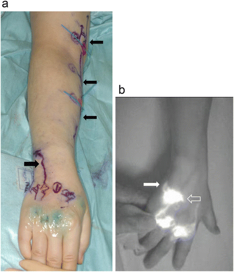
Fig. 43.4
ICG fluorescence lymphography in the right arm of a 47-year-old woman with breast cancer-related lymphedema
(a) Perioperative ICG fluorescence lymphography shows linear fluorescence (closed arrows) in the hand and arm. Five LVSEAs were performed in the upper limb
(b) Postoperative ICG fluorescence lymphography performed 7 months after surgery. Dermal backflow (open arrow) that was not observed at the operation has developed at the site of LVSEA in the hand. Linear fluorescence (closed arrow) that was likewise not apparent at the operation is observed from dermal backflow to the forearm
43.3 Case Presentation
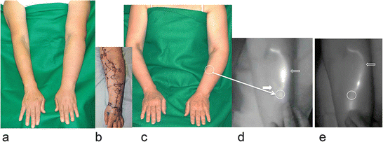
43.3.1 Mid-term Results (Fig. 43.5)
A 56-year-old woman with left upper-limb lymphedema after breast cancer treatment was referred for lymphatic microsurgery. This patient received complex decongestive physiotherapy for 3 years, but edema in the forearm (volume, 1880 ml) had not improved (Fig. 43.5a). LVSEA was planned and three anastomoses in the upper limb were performed (Fig. 43.5b). Volume of the left upper limb was decreased immediately after surgery and good condition was maintained with a volume of 1646 ml at 2 years and 10 months after surgery (Fig. 43.5c). ICG fluorescence lymphography showed one anastomosis was patent, one was undetectable, and one was non-patent at both 6 months (Fig. 43.5d) and 2 years 10 months (Fig. 43.5e) after surgery.
 ICG Fluorescent Image-Guided Surgery in Head and Neck Cancer
 Regional Lymph Node Dissection Assisted by Indocyanine Green Fluorescence Lymphography and Angiography for Stage III Melanoma
 Fluorescence Imaging for Intraoperative Identification of Pancreatic Leak
 Intraoperative Detection of Hepatocellular Carcinoma Using Indocyanine Green Fluorescence Imaging
 Indocyanine Green Fluorescent Lymphography and Microsurgical Lymphaticovenous Anastomosis
 Detection of Hepatic Micrometastases from Pancreatic Cancer


Related posts:
ICG Fluorescent Image-Guided Surgery in Head and Neck Cancer
Regional Lymph Node Dissection Assisted by Indocyanine Green Fluorescence Lymphography and Angiography for Stage III Melanoma
Fluorescence Imaging for Intraoperative Identification of Pancreatic Leak
Intraoperative Detection of Hepatocellular Carcinoma Using Indocyanine Green Fluorescence Imaging
Indocyanine Green Fluorescent Lymphography and Microsurgical Lymphaticovenous Anastomosis
Detection of Hepatic Micrometastases from Pancreatic Cancer
Stay updated, free articles. Join our Telegram channel
Full access? Get Clinical Tree
