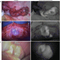
Fig. 20.1
Some linear enhancement from each of the perforators is observed in the TRAM flap
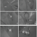
The TRAM flap is enhanced from Zone I diffusely, when the movement of the line stops (Fig. 20.2: Zone Classification of a TRAM Flap). On the ipsilateral side of the vascular pedicle, the diffuse enhancement extends toward the Zone III, and in most of the cases, the whole of the Zone III is enhanced. On the contralateral side of the vascular pedicle, the diffuse enhancement extends beyond the center line, and the enhancement stops between Zone II and the lateral edge of Zone IV (Fig. 20.3).
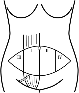
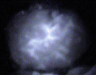
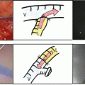
Fig. 20.2
Zone classification of the TRAM flap
Fig. 20.3
The TRAM flap is enhanced diffusely
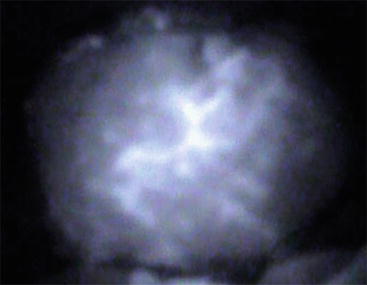
Diffuse enhancement starts and stretches between the linear enhancement. And then, the diffuse enhancement stagnates in 1–2 s (Fig. 20.4).
The enhanced area starts to spread laterally again, after the movement of the enhanced region stops or move very slowly in a few minutes. But the movement of the enhanced region after the stagnation is slower than it was before the stagnation. This extension moves slower than the former extension. The enhanced region spreads slowly toward the lateral edge of the TRAM flap and the whole region is eventually enhanced in most flaps. But, in some cases, the spread of the enhancement stops between Zones II and IV of the flap (Figs. 20.5 and 20.6).
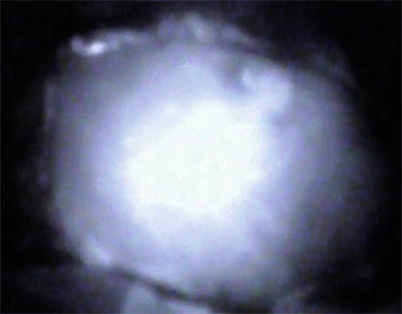
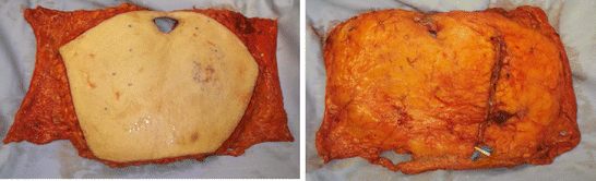
Fig. 20.5
Thirty seconds after Fig. 20.4, enhanced region extends and almost all of the region of the TRAM flap is enhanced
Fig. 20.6
Harvested TRAM flap. Left: Skin surface, Right: Fat surface with vascular pedicle
20.3 Evaluation
Eventually, the whole region of the flap becomes enhanced in most TRAM flaps. Nevertheless, partial fat necrosis and partial flap loss could still occur. The usefulness of this process depends on the proper evaluation of the effect of the enhancement with ICG. The most effective evaluation is done at the time when the initial diffusion stops or becomes stagnant.
20.4 Results
When only grafting regions which were enhanced before stagnation occurred, there were no complications of the TRAM flaps such as partial fat necrosis or partial flap loss. However, of the 17 cases in which grafts included non-enhanced regions, seven showed partial fat necrosis (Fig. 20.7).
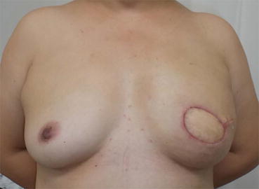
Fig. 20.7
Left reconstructed breast shows symmetric shape without partial fat necrosis
In cases of patients who had mid-abdominal scars, if the patient had a complete midline scar from the cephalad edge to caudal edge, there was no case of the flap in which contralateral region of the vascular pedicle was enhanced. However, the flaps had intact areas a few centimeters from the umbilicus, and the contralateral region of the flap was enhanced in some cases. There was a tendency for the contralateral side of the flap to be enhanced, when the patient had a perforator near the intact area of the midline.
20.5 Discussion
It is useful for the plastic surgeon to use ICG (indocyanine green) for the breast reconstruction surgery, as ICG is also usually used in breast cancer surgery for detecting sentinel lymph nodes.
In the plastic and reconstructive surgery field, Eren et al. [1] describe that ICG fluorescence angiography was useful to evaluate the reliable circulation of the axial pattern flap in rat models. The first clinical report of evaluation of the circulation of the flap with ICG was reported by Still et al. [2] In this report, 18 patients who were burned underwent ICG fluorescence imaging for the evaluation of the circulation of the flap. In one patient, two flaps were evaluated. The entire flaps were enhanced and the whole region survived in 16 flaps. However, in another flap, almost the entire area of the flap was not enhanced and in the end that flap showed almost complete necrosis. In two flaps, un-enhanced regions survived. Holm et al. [3] described the usefulness of ICG fluorescence enhancement for evaluating which regions would survive in 11 axial pattern flaps and four random pattern flaps. Afterward, ICG fluorescence angiography was used to evaluate the free flaps [4, 5].
Related posts:
 ICG Fluorescent Image-Guided Surgery in Head and Neck Cancer
 Regional Lymph Node Dissection Assisted by Indocyanine Green Fluorescence Lymphography and Angiography for Stage III Melanoma
 Fluorescence Imaging for Intraoperative Identification of Pancreatic Leak
 Intraoperative Detection of Hepatocellular Carcinoma Using Indocyanine Green Fluorescence Imaging
 Indocyanine Green Fluorescent Lymphography and Microsurgical Lymphaticovenous Anastomosis
 Principle and Development of ICG Method
Stay updated, free articles. Join our Telegram channel

Full access? Get Clinical Tree


