• Describe the process of radiographic image formation. • Explain the process of beam attenuation. • Identify the factors that affect beam attenuation. • Describe the x-ray interactions termed photoelectric effect and Compton effect. • State the composition of exit radiation. • State the effect of scatter radiation on the radiographic image. • Explain the process of creating the various shades of image densities and brightness. • Differentiate among conventional and digital imaging. • Define fluoroscopy and describe the process of image intensification. The term differential is used because varying anatomic parts do not absorb the primary beam to the same degree. Anatomic parts composed of bone absorb more x-ray photons than parts filled with air. Differential absorption of the primary x-ray beam creates an image that structurally represents the anatomic area of interest (Figure 8-1). As the primary x-ray beam passes through anatomic tissue, it loses some of its energy. With the photoelectric effect, the ionized atom has a vacancy, or electron hole, in its inner shell. An electron from an outer-shell drops down to fill the vacancy. Because of the difference in binding energies between the two electron shells, a secondary x-ray photon is emitted (Figure 8-2). This secondary x-ray photon typically has very low energy and is therefore unlikely to exit the patient. Some incoming photons are not absorbed, but instead lose energy during interactions with the atoms composing the tissue. This process is called scattering. It results from the diagnostic x-ray interaction with matter known as the Compton effect. The loss of some energy of the incoming photon occurs when it ejects an outer-shell electron from a tissue atom. The ejected electron is called a Compton electron or secondary electron. The remaining lower-energy x-ray photon changes direction and may leave the anatomic part to interact with the image receptor (Figure 8-3).
Image Production
Differential Absorption
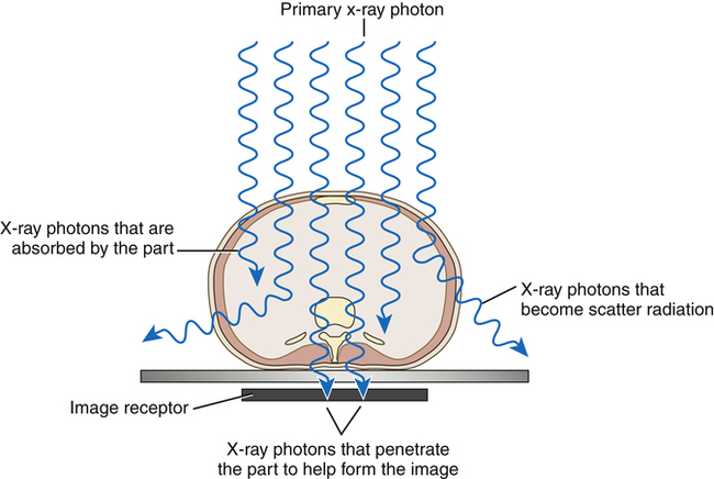
As the primary beam interacts with the anatomic part, x-ray photons are transmitted, absorbed, and scattered based on the tissue’s composition. Differential absorption of the primary x-ray beam creates an image that structurally represents the anatomic area of interest.
Beam Attenuation
Absorption
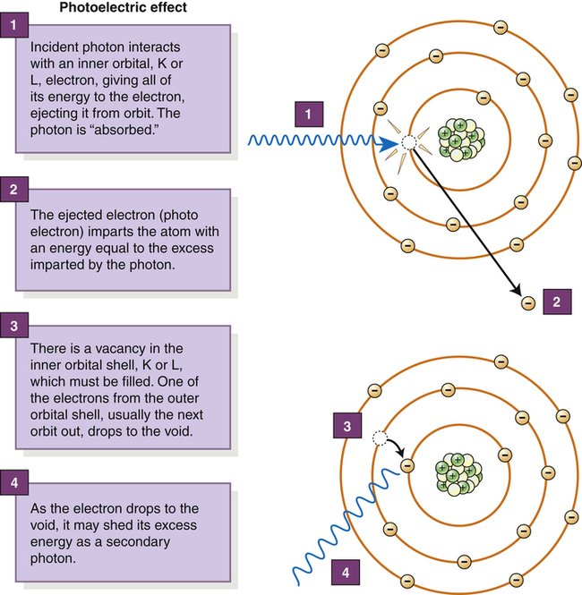
The photoelectric effect is responsible for total absorption of the incoming x-ray photon.
Scattering
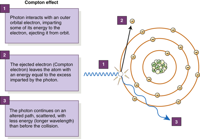
During the Compton effect, the incoming x-ray photon loses energy and changes its direction.
![]()
Stay updated, free articles. Join our Telegram channel

Full access? Get Clinical Tree


Image Production
 Make the Physics Connection 8-1
 Critical Concept 8-1
 Make the Physics Connection 8-2
 Critical Concept 8-2
 Critical Concept 8-3