Fig. 1
A schematic visualization of the tympanic cavity as a box after removal of the posterior wall with tympanic membrane (1) and malleus (2) on the lateral wall, incus (3), Eustachian tube (4) and tensor tympani tendon (5) at the level of the cochleariform process on the anterior wall, stapes (6) at the oval window (7) on the medial wall, pyramidal eminence (8) with stapedius tendon (9) attached to the neck of the stapes, jugular bulb (10), round window (11), cochlear promontory (12) with cochlea (13) and semicircular canals (14) shining trough the medial wall of the tympanic cavity
The tympanic cavity can be divided into three subdivisions in the coronal plane: the epi-, meso-, and hypotympanum. The epitympanum is the attic, formed by an imaginary line drawn between scutum and tympanic segment of the facial nerve. The mesotympanum is the middle part delineated by the line between scutum and tympanic segment of the facial nerve on one hand, and a line connecting the tympanic annulus and the base of the cochlear promontoryon the other hand. The hypotympanum is the lowest part of the tympanic cavity and lies below the line between tympanic annulus and the base of the cochlear promontory.
1.2.2 Auditory Ossicles
The tympanic cavity contains three auditory ossicles: the malleus, the incus, and the stapes (Fig. 2). The ossicles act as a lever transforming the large and weak motion of the tympanic membrane into a small and forceful movement of the stapes. The malleus is attached to the tympanic membrane and the stapes is connected to the fenestra vestibuli, traditionally referred to as the oval window. In between the incus is suspended by delicate articulations, the incudomalleal and incudostapedial joints.
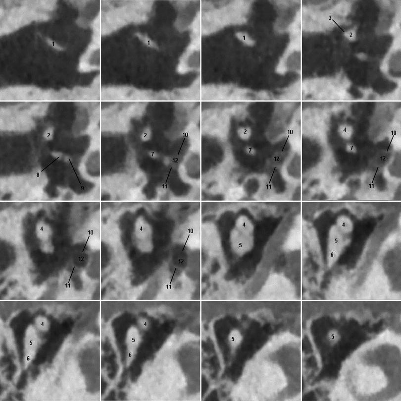
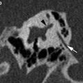
Fig. 2
Consecutive axial 0.3 mm thick CBCT images through a normal middle ear showing the ossicular chain with malleus manubrium (1), neck (2), lateral process (3) and head (4); incus body (5) with short (6) and long process (7) and its lenticular process (8) articulating with the head of the stapes (9); anterior (10) and posterior crus (11) of the stapes and the foramen obturatorium (12)
The malleus, named after the resemblance to a hammer, consists of a head, neck, and three processes:, the manubrium, the anterior and lateral processes.
The incus resembling an anvil consists of a corpus and 2 crura (processes). The anterior part of the corpus articulates with the head of the malleus in a saddle-shaped diarthrosis. The two processes are positioned perpendicular to each other with the short process running almost horizontally backward and the long process descending nearly vertically and bending medially, ending in a little notch covered by cartilage, the lenticular process, articulating with the head of the stapes.
The stapes similar to a stirrup, consists of a head, neck, 2 crura and a base. In between the 2 crura lies the foramen obturatorium. The 2 crura diverge from the neck and are at their end connected to an oval plate that is fixed to the fenestra vestibuli by a ring of ligamentous fibers, the annular ligament.
1.2.3 Suspensory Apparatus
The malleus, incus, and stapes are situated between the tympanic membrane and oval window, and are supported by the tympanic membrane, the anterior, superior, and lateral malleal ligaments, the posterior incudal ligament, the tendons of the tensor tympani and stapedius muscles, and the annular ligament (Fig. 3).
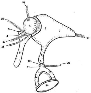
Fig. 3
A schematic visualization of the ossicular chain and its suspensory apparatus with malleus and its head (1), neck (2), lateral (3) and anterior process (4) and manubrium (5); incus with body (6), short (7) and long process (8) ending in the lenticular process (9); stapes with head (10), neck (11), anterior (12) and posterior crus (13), and footplate (14). The superior malleal ligament (15) inserts on the malleal head (1); the lateral malleal ligament (16), anterior malleal ligament (17), and tensor tympani tendon (18) insert on the malleal neck (2); the posterior incudal ligament (19) inserts on the incus short process (7); and the stapedius muscle tendon (19) mostly inserts on the stapes neck (11)
The malleus is supported by the superior, anterior, and lateral malleal ligaments, the tensor tympani muscle tendon, the tympanic membrane, and the incudomalleal joint. The incus is supported by the posterior incudal ligament and two joints, the incudomalleal and incudostapedial joints. The stapes is supported by the stapedius muscle tendon and the incudostapedial joint.
Axial CBCT images are excellent to see the anterior malleal ligament (AML), posterior incudal ligament (PIL), and the stapedius muscle tendon. Coronal images are excellent to see the superior malleal ligament (SML) and the lateral malleal ligament (LML). The tensor tympani muscle tendon can be appreciated on both axial and coronal images (Table 1).
Table 1
Origin and insertion of suspensory middle ear ligaments and tendons
ORIGIN → INSERTION | |
|---|---|
Axial | |
AML | Anterior epitympanic wall → malleus neck |
PIL | Incudal fossa → incus short process |
Stapedius tendon | Pyramidal eminence → stapes neck |
Tensor tympani tendon | Cochleariform process → malleus neck |
Coronal | |
SML | Epitympanic roof → malleus head |
LML | Notch of rivinus → malleus neck |
Tensor tympani Tendon | Cochleariform process → malleus neck |
The AML is attached at one end to the malleal neck, just above the anterior process, and to the anterior tympanic wall by the other end. The PIL connects the incus short process to the incudal fossa (Fig. 4). The SML runs from the roof of the epitympanic recess to the malleus head (Fig. 5). The LML extends from the posterior part of the notch of Rivinus to the malleus neck (Fig. 6).
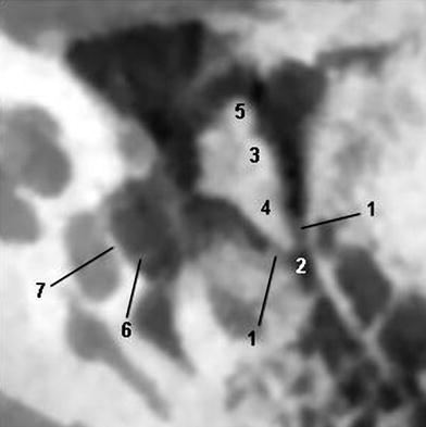
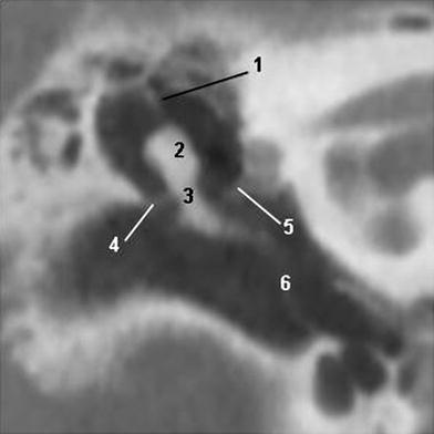
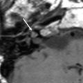
Fig. 4
Axial 0.3 mm thick CBCT image through a normal middle ear showing the posterior incudal ligament (1), fossa incudis (2), incus body (3) and its short process (4), malleus head (5), crus posterior of the stapes (6), and foramen ovale (7)
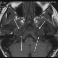
Fig. 5


Coronal 1 mm thick CBCT image through a normal middle ear showing the superior malleal ligament (1), malleus head (2), malleus neck (3), lateral malleal ligament (4), tensor tympani tendon (5), and tympanic membrane (6)
Related posts:


Stay updated, free articles. Join our Telegram channel
Full access? Get Clinical Tree
