Immunotherapy in head and neck cancer introduces novel tumor response patterns and immune-related toxicities that can mimic disease progression and challenge traditional imaging assessment. Accurate interpretation requires in-depth understanding of immune checkpoint inhibitors, chimeric antigen receptor-T cell therapy, and oncolytic virus treatments, along with recognition of atypical responses such as pseudoprogression, hyperprogression, dissociated response, and durable response. Incorporating modified imaging criteria like iRECIST and imPERCIST enhances diagnostic accuracy. Radiologists play a vital role in identifying immune-related adverse events across organ systems, guiding appropriate management, and supporting continued treatment when effective.
Key points
-
•
Immunotherapy is reshaping head and neck cancer care, introducing atypical tumor response patterns and immune-related toxicities.
-
•
Radiologists must understand mechanisms of immune therapies and recognize unique imaging features.
-
•
Modified response criteria (iRECIST and imPERCIST) help distinguish true progression from pseudoprogression.
-
•
Imaging plays a key role in detecting immune-related adverse events and guiding management.
Abbreviations
| APC | antigen-presenting cell |
| ASTCT | American Society for Transplantation and Cellular Therapy |
| CAR | chimeric antigen receptor |
| CR | complete response |
| CRS | cytokine release syndrome |
| CTCAE | Common Terminology Criteria for Adverse Events |
| CTLA-4 | cytotoxic T-lymphocyte antigen-4 |
| EGFR | epithelial growth factor receptor |
| GM-CSF | granulocyte macrophage colony-stimulating factor |
| HLH | hemophagocytic lymphohistiocytosis |
| HNSCC | head and neck squamous cell carcinoma |
| ICANS | immune effector cell-associated neurotoxicity syndrome |
| ICI | immune checkpoint inhibitor |
| iCPD | immune-confirmed progressive disease |
| imPERCIST | immunotherapy-modified PET Response Criteria in Solid Tumors |
| irAE | immune-related adverse event |
| iRESIST | immune Response Evaluation Criteria in Solid Tumors |
| iUPD | immune unconfirmed progressive disease |
| NK | natural killer |
| PD | progressive disease |
| PD-1 | programmed death-1 |
| PD-L1 | programmed death-ligand 1 |
| PERCIST | PET Response Criteria in Solid Tumors |
| PFS | progression-free survival |
| PMD | progressive metabolic disease |
| PNS | perineural spread |
| PR | partial response |
| RESIST | Response Evaluation Criteria in Solid Tumors |
| SD | stable disease |
| TKI | tyrosine kinase inhibitor |
| Treg | Regulatory T |
| T-VEC | talimogene laherparepvec |
Introduction
The use of immunotherapy has revolutionized cancer treatment and improved survival in patients over the past decade, including in head and neck oncology. , Immune-based therapies, such as immune checkpoint inhibitors (ICIs), have changed standard-of-care regimens. For example, in recurrent or metastatic head and neck squamous cell carcinoma (HNSCC), ICIs like pembrolizumab and nivolumab have demonstrated significant survival benefits and are now the first-line or second-line treatments. With this paradigm shift, radiologists must understand the mechanisms, distinct response patterns, and toxicity that accompany these treatments. Unlike traditional, cytotoxic chemotherapy, immunotherapy can produce delayed or atypical tumor responses. Therefore, familiarization with immune-related response criteria and imaging appearances of immune side effects is critical to avoid misinterpretation or premature discontinuation of an effective therapy. This article summarizes key immunotherapy mechanisms (including targeted antibodies, chimeric antigen receptor [CAR]-T cells, and oncolytic virus therapy), current applications in head and neck cancer, response assessment guidelines, atypical response patterns, and common immune-related adverse events (irAEs) pertinent to radiologists.
Mechanism of action of targeted antibodies
Small Molecules Versus Monoclonal Antibodies
Targeted cancer therapies include small molecule drugs and monoclonal antibodies, which differ in structure and mechanism. Small molecule inhibitors are low molecular weight (0.5–1 kDa) compounds produced by chemical synthesis and often have the suffix “-ib” (eg, erlotinib), whereas monoclonal antibodies are large (∼150 kDa) bioengineered immunoglobulin proteins and end in “-mab” (eg, cetuximab). Due to their size, small molecules can often penetrate cell membranes and bind individual proteins, affecting intracellular signaling pathways; they are ideal for targeting enzymes or receptors with small, well-defined active sites. Monoclonal antibodies, on the other hand, are generally restricted to extracellular targets such as cell surface receptors or ligands; they are highly specific and bind a single target with high affinity.
Tyrosine Kinase Inhibitors: B-raf proto-oncogene and Mitogen-activated protein kinase kinase Inhibitors in V600E Mutated Tumors
Tyrosine kinase inhibitors (TKIs) are small molecules that competitively inhibit the adenosine triphosphate binding sites of specific tyrosine kinase enzymes, thereby blocking downstream signaling in the growth and survival pathways of cancer cells. Many TKIs target dysregulated pathways common in head and neck malignancies. B-raf proto-oncogene (BRAF) and mitogen-activated protein kinase kinase (MEK) inhibitors form a notable subset of TKIs and target the mitogen-activated protein (MAP) kinase pathway. BRAF inhibitors specifically target the BRAF protein, a serine/threonine-protein kinase involved in cell signaling. , Mutations in the BRAF gene, especially the V600 mutations, are common in certain thyroid cancers and cutaneous melanoma (for instance ∼50% of cutaneous melanomas and some thyroid cancers). These mutations drive aberrant activation of the BRAF-MEK-extracellular signal-regulated (ERK) pathway leading to uncontrolled cell growth. On the other hand, MEK inhibitors target the MEK1 and MEK2 proteins, which are downstream of BRAF in the MAP kinase pathway. Combined BRAF and MEK inhibition (eg, dabrafenib plus trametinib) can induce dramatic tumor regressions in BRAF V600 mutated tumors and has become an established therapy for BRAF-mutant melanoma and anaplastic thyroid cancer ( Fig. 1 ).
Mechanism of action of a BRAF and MEK inhibitor.
Epithelial Growth Factor Receptor and CD20 Monoclonal Antibodies
Epithelial growth factor receptor (EGFR) is a protein on the surface of cells that, when overexpressed or mutated, can lead to uncontrolled cell divisions, especially in cancers like lung, colorectal, and head and neck cancers. EGFR inhibitors (eg, cetuximab) target this receptor to inhibit tumor growth, as it is overexpressed in the majority of HNSCCs.
The CD20 antigen is a protein found on the surface of B cells monoclonal antibodies. CD20-targeted monoclonal antibodies (eg, rituximab) are a mainstay in the treatment of B cell malignancies (eg, non-Hodgkin lymphoma and chronic lymphocytic leukemia), including those arising in the head and neck region, as they induce cell death and recruit immune cells to attack CD20-expressing cells ( Fig. 2 ).
Mechanism of action of a CD20 monoclonal antibody (rituximab). Binding of rituximab activates the complement cascade, which generate the membrane attack complex that lyses B cells. Complement activation also leads to phagocytosis by macrophage recognition of complement factor and Fcy receptor. Binding of rituximab also allows interaction with NK cell.
Mechanism of action of immune checkpoint inhibitors
Cytotoxic T-lymphocyte Antigen-4 Inhibitors
The cytotoxic T-lymphocyte antigen-4 (CTLA-4) is an inhibitory receptor expressed on T cells (particularly regulatory and activated T cells) that downregulates T cell activation early in the immune response. CTLA-4 competes with the co-stimulatory receptor CD28 for binding B7 ligands (CD80/CD86) on antigen-presenting cells (APCs), thereby dampening T cell priming in lymphoid tissue, and effectively acting as a “brake” on T cell proliferation and function ( Fig. 3 A, B). Tumors can exploit this pathway to reduce antitumor immunity. CTLA-4 inhibitors, such as ipilimumab which was approved as first line for melanoma, block CTLA-4, allowing sustained T cell activation against tumor antigens (see Fig. 3 B). However, in other head and neck cancers, due to less tumor shrinkage and more serious side effects, CTLA-4 inhibitors are not first-line agents and are being studied in combination with other therapies.
( A ) CTLA-4 pathway. Regulatory T (Treg) cells inhibit APCs by 3 main mechanisms: (1) Depleting immune-stimulating cytokines; (2) producing immunosuppressive cytokines; and (3) constitutively expressing CTLA-4, which blocks the priming and activation of naïve CD4 + T (Tconv) cells to APCs. ( B ) Mode of action of CTLA-4 inhibitors. T cell activation requires 2 signals; signal 1 between MHC and T cell receptor and signal 2 between CD80/86 molecules on the antigen receptor cell (APC) and CD28 on the T cell. Activation leads to CTLA-4 upregulation and translocation to the cell surface, which competitively binds to CD80/86 leading to downregulation and T cell inactivation. Ipilimumab (Ipi) blocks CTLA-4 and CD80/86 binding, allowing for ongoing activation of T cells and antitumor effect.
Programmed Death-1 and Programmed Death-Ligand 1 Inhibitors
The programmed death-1 (PD-1) receptor on T cells and its ligand PD-L1 represent another immune checkpoint pathway that predominantly functions in peripheral tissues to limit active immune responses. PD-1 is an inhibitory receptor that, when bound to PD-L1, transmits a negative signal that reduces T cell proliferation and cytotoxic function. It also causes B cell inhibition and natural killer (NK) cell dysfunction, and among other effects to dial down immune activation. Many head and neck tumors upregulate PD-L1 as an immune evasion mechanism ( Fig. 4 A). Monoclonal antibodies targeting PD-1 (such as nivolumab and pembrolizumab) or PD-L1 (such as durvalumab and atezolizumab) block this interaction and reverse the tumor suppressive effect on anticancer immune response ( Fig. 4 B).
( A ) PD-1/PD-L1 pathway. When overexpressed PD-L1 on tumor cells combines with PD-1 on cytotoxic T cells, an inhibitory second signal transmits to T cells, causing effector T cells exhaustion and dysfunction. PD-L1 also binds to PD-1 on Tregs, resulting in immune suppression by raising the threshold for T cell activation. The overexpressed tumor PD-L1 binding also leads to dysfunction of NK cell and inhibits the proliferation of CD4 + and CD8 T+ cells. ( B ) Mode of action of PD-1 and PD-L1 inhibitors. PD-1 antibodies can competitively inhibit the binding of PD-1 to PD-L1, while PD-L1 antibodies inhibit the binding to PD-1. Both inhibit the activation of the PD-1/PD-L1 signal pathway and reverse the suppressive effect.
Mechanism of action of chimeric antigen receptor-T cell and talimogene laherparepvec therapy
Chimeric Antigen Receptor-T Cell Therapy
CAR-T cell therapy is a form of adoptive cellular immunotherapy that involves reprogramming a patient’s own T lymphocytes to recognize and attack cancer cells. The process involves extracting the patient’s T cells via leukapheresis, then genetically engineering them to express a synthetic CAR that targets a specific tumor-associated antigen. These receptors typically combine an antigen-binding domain (often derived from an antibody) that targets specific proteins on cancer cells, and a signaling domain that activates the T cell once it binds to the cancer cell. Once engineered, the CAR-T cells are reinfused back into the patient, usually following a lymphodepleting chemotherapy regimen to enhance T cell engraftment ( Fig. 5 ). The most successful applications of CAR-T therapy to date have been in CD19-positive B cell malignancies (leukemias and lymphomas), where anti-CD19 CAR-T cells can induce high remission rates.
CAR-T cell therapy.
Talimogene Laherparepvec Therapy
Oncolytic virus therapy uses replication-competent viruses to infect and kill cancer cells and stimulate antitumor immunity. Talimogene laherparepvec (T-VEC) is the first Food and Drug Administration-approved oncolytic virus therapy, indicated for advanced melanoma, and it serves as a prototype for this class. T-VEC is a genetically modified herpes simplex virus type 1 engineered to selectively replicate within tumor cells and to express the cytokine granulocyte macrophage colony-stimulating factor (GM-CSF). After intratumoral injection, T-VEC infects and lyses tumor cells, causing the release of tumor antigens. The expression of GM-CSF by the virus helps recruit and mature dendritic cells to the tumor site, enhancing antigen presentation and priming of T cells against tumor antigens. Through these mechanisms, T-VEC can induce not only regression of the injected lesions, but also polyclonal immune-mediated regression of distant metastases in some cases ( Fig. 6 ).
Mode of action of T-VEC. GM-CSF. Tumor-deprived antigens.
Response evaluation guidelines
Assessing tumor response to immunotherapy requires modified imaging criteria, as traditional criteria like Response Evaluation Criteria in Solid Tumors (RECIST) 1.1 may not adequately capture the unique patterns of response. Two main domains are considered: anatomic response (typically measured on computed tomography [CT]/MR imaging) and metabolic response (measured on PET). For each, immunotherapy-tailored criteria have been developed.
Response Evaluation Criteria in Solid Tumors 1.1 Versus Immune Response Evaluation Criteria in Solid Tumors
RECIST 1.1 is the conventional set of criteria used to categorize tumor response on anatomic imaging. RECIST 1.1 defines complete response (CR) as disappearance of target lesions, partial response (PR) as at least a 30% decrease in the sum of diameters of target lesions, progressive disease (PD) as at least 20% increase in the sum of diameters (with a minimum 5 mm absolute increase) or the appearance of new lesions, and stable disease (SD) as changes that fall between PR and PD thresholds. While RECIST 1.1 has been the cornerstone for cytotoxic chemotherapy trials, it can be misleading for immunotherapy. This is because immunotherapeutic agents can cause atypical response patterns like pseudoprogression, where tumors initially enlarge due to immune cell infiltration or transient inflammation before later shrinking. Under RECIST 1.1, the initial enlargement or new lesions would be labeled PD, potentially leading to an incorrect cessation of therapy.
To address this, immune RECIST (iRECIST) guidelines were introduced in 2017 as an adaptation of RECIST 1.1 for trials of immunotherapeutics. iRECIST retains the RECIST 1.1 measurement principles but adds new categories to account for unconfirmed progression. When imaging shows findings meeting PD criteria by RECIST in a patient on immunotherapy, iRECIST designates this as immune unconfirmed progressive disease (iUPD) rather than immediate PD. The patient can continue therapy, and a follow-up scan is obtained (usually 4–8 weeks later) to confirm or refute progression. If the follow-up scan shows continued tumor growth or additional new lesions, then immune-confirmed PD (iCPD) is assigned, confirming progression. However, if the follow-up scan shows tumor stabilization or shrinkage, the initial changes are deemed pseudoprogression and the response category may be modified back to immune SD or even immune PR, and treatment can be continued. Essentially, iRECIST mandates confirmation of progression on a subsequent imaging examination (in the absence of rapid clinical deterioration) before labeling a patient as having PD. This approach acknowledges that some patients who appear to worsen radiographically at first may later respond. It is important to note that iRECIST does not require adding the measurements of new lesions to the tumor burden sum, whereas RECIST 1.1 would count new lesions as progression regardless of size. For routine clinical practice, formal iRECIST application may not always be done, but radiologists can apply the same principles by suggesting confirmation when radiologic progression is suspected but the patient’s clinical status is stable or improving.
PET Response Criteria in Solid Tumors 1.0 Versus Immunotherapy-modified PET Response Criteria in Solid Tumors
In metabolic imaging with 18F-Fluorodeoxyglucose (FDG) PET, PERCIST 1.1 (PET Response Criteria in Solid Tumors) is an analog to RECIST that evaluates treatment response based on changes in FDG uptake rather than size. PERCIST 1.0 typically assess standardized uptake value normalized to lean body mass (SUL) peak of up to the 5 hottest lesions (usually one lesion per organ) and defines metabolic response criteria. For example, a complete metabolic response requires resolution of uptake in target lesions to background level, and progressive metabolic disease (PMD) is defined by a significant increase in uptake (>30% in SUL peak) or new FDG-avid lesions appearing. Under baseline PERCIST, the appearance of a new FDG-avid lesion suspicious for tumor is considered PMD (analogous to PD in RECIST).
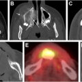
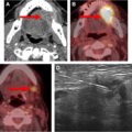
Immunotherapy can confound PET interpretation because immune activation can result in increased uptake at sites of preexisting metabolically active disease, as well as causing new FDG avid foci secondary to recruitment of immune cells to sites of previously PET occult microscopic malignant disease, or secondary to a benign immune response and/or irAE. Hence, an immunotherapy-modified PERCIST (imPERCIST) has been proposed to avoid mischaracterizing progression in this setting. Like PERCIST, imPERCIST quantifies the SUL peak in up to 5 lesions to track changes in metabolic activity. The key difference is that imPERCIST does not automatically label new or increasing FDG-avid lesions as progression if the patient is clinically stable. Instead, new and increasing lesions on a PET at 8 weeks following treatment initiation are labeled unconfirmed PMD (similar to iRECIST’s iUPD), triggering a short-term follow-up PET 4 weeks later to evaluate the stability of these findings. If the new and increased lesions show continuing increase in uptake above that seen at the 8-week PET, the results are characterized as confirmed PMD. If instead the new and increased lesions show stable or decreased uptake relative to that seen on the 8-week PET, the results are downgraded and characterized as stable metabolic disease or partial metabolic response, respectively. imPERCIST has been suggested to correlate better with overall survival in immunotherapy patients compared with PERCIST. By integrating imPERCIST principles, radiologists and nuclear medicine physicians can avoid calling PMD prematurely.
Related posts:
Stay updated, free articles. Join our Telegram channel

Full access? Get Clinical Tree


