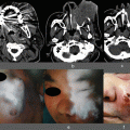
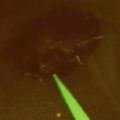
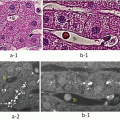
Fig. 16.1
The mechanism for detecting ICG-induced fluorescence by NIR camera system
Once binding with plasma protein, ICG can emit fluorescence of maximum wavelength around 845 nm when it is excited by near-infrared light wavelength of 750–810 nm. This fluorescence of ICG can be detected at least 10 mm beneath the skin surface by infrared camera unit. This camera system has a NIR camera, which has a cutoff filter wavelength below 830 nm, in the center to capture the fluorescence from lymphatic tracts, surrounded by many light-emitting diode (LED) units of wavelength 760 nm to excite protein-binding ICG
16.2 Objectives
SLNB using ICG-NIR method can be applied for melanoma as well as for various types of nonmelanoma skin cancers tend to lead to lymphatic metastases, e.g., MCC, EMPD, SCC, and porocarcinoma; although the efficacy of SLNB for nonmelanoma skin cancers has not been well proven. For SLNB, patients who exhibited clinically- or radiographically- obvious metastasis of the regional lymph nodes or other organs must be excluded. However, in patients with tumors in the midline of the trunk, who are diagnosed as having hemilateral regional lymph node metastases, clinical stage can be evaluated using the contralateral SLNs.
16.3 Methods
16.3.1 Administration of ICG
As a tracer, 25 mg of ICG was diluted into 5 ml of sterile distilled water. The concentration of ICG was reduced based upon the distance between the injection sites and the SLNs. Up to 1.0 ml of this solution was injected intracutaneously into several locations around the tumor. The desirable points for tracer administration in skin cancers, other than some cases of EMPD, are in the vicinity of the tumor or the scar following excisional biopsy (Fig. 16.2a). For EMPD, the sites for tracer administration should be carefully determined according to the size and surface configuration of the plaque. For flattened small lesions (less than 5 cm in maximum diameter), the tracer should be injected nearby. For large plaques (over 5 cm in maximum diameter), the tracer can be injected in the proximity of the most elevated or eroded lesions, which suggest histopathologic intradermal cancer invasion, within or besides the plaque (Fig. 16.3a and c). In order to reduce the pain of intracutaneous injection, local anesthetic tape may be pasted at the estimated injection sites one hour prior to administration. During intradermal injections of ICG, oozing ICG should be absorbed or wiped off immediately to avoid unnecessary illumination of the skin surface and operative field. Surgical gloves that become contaminated with ICG should be changed for the same reason. Another type of vital dye, i.e. 0.4 % (w/v) indigo carmine or 2 % isosulfan blue could be administered several minutes prior to ICG injection in order to support the blue staining of the lymphatic tracts.
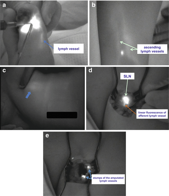
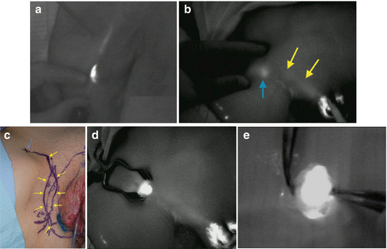

Fig. 16.2
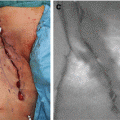
(a) Linear fluorescence of lymph tract appeared shortly after intradermal injection of ICG. (b) Fluorescence of ascending lymph vessels observed on the lower leg (light-green arrows). (c) Within 10 min, fluorescence from inguinal node (blue arrow) becomes visible from the surface. (d) After incision, round fluorescence of SLN and linear fluorescence of afferent lymph vessels (red arrow) become visible. (e) Stumps of the amputated lymph vessels can be observed after SLN dissection

Fig. 16.3
(a) Linear fluorescence of lymph tract appeared during intradermal injection of ICG. (b) Fluorescence of ascending lymph vessels (yellow arrows) and fluorescence from inguinal node (blue arrow) become visible within several minutes after ICG administration. (c) Clinical feature of detected lymph tracts (yellow arrows) to SLN (blue arrow). (d) After incision, round fluorescence of SLN becomes visible. (e) Resected SLN still generates fluorescence
16.3.2 Observation of the Lymph Tracts (Fig. 16.1)
Immediately after injection of the ICG, fluorescence of the lymphatic channels to the SLNs can be observed from the skin surface (Figs. 16.2b–c and 16.3b–c) using the infrared camera system [PDE (Photodynamic Eye) or PDE-neo, Hamamatsu Photonics K.K., Hamamatsu, Japan] under normal room light. Only the operating lamps needed to be switched off for the ICG-INR guided observation. The required time for detecting fluorescence of SLNs is within 5 min for inguinal nodes. For the trunk, head, neck, and upper limbs, a rather longer detecting time tends to be necessary. Diffuse fluorescence of the skin overlying the regional lymph nodes (Fig. 16.4), which reflects the dermal backflow and pooling of tracer caused by lymphatic obstruction, sometimes suggests the existence of regional node metastasis. In the axillary region, observation of the fluorescence from the SLNs is more difficult than the inguinal area because of the deeper location of the SLNs covered by thicker fat tissues. In such cases, the overlying skin and subcutaneous tissue should be pressed in order to reduce the distance from the skin surface to the SLNs.
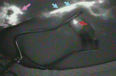

Fig. 16.4
Dermal backflow observed at left inguinal region of EMPD patient. SLN could not be observed from the surface because it was masked by the brighter fluorescent pooling of the skin (blue arrows), which is called “dermal backflow.” SLN (red arrow) could be visible only after skin incision was made. Pink arrow shows the ICG-injected genital site
16.3.3 Surgical Resection of SLNs
Detected SLNs should be removed surgically (Fig. 16.3d) for histological examination. After making the incision, the linear fluorescence of the afferent lymph vessels to SLNs is observed (Fig. 16.2d). To avoid diffuse illumination that can interfere with observation of the lymphatic flow and the SLNs, surgery for lymph node detection must be performed carefully. The detected lymph vessels (Fig. 16.2d) must be carefully ligated or cauterized to avoid the leakage of the ICG that led to the disseminated fluorescence at the biopsy basin and disturbed the further detections of other SLNs. This series of lymph node dissections should be completed as quickly as possible within about 30 minutes to avoid the spread of the fluorescence to second-tier lymph nodes. After completion of the dissection of the SLNs, residual lymph nodes must be examined by gamma-prove and the NIR camera system. At this time, punctate illumination, which suggests the stumps of amputated lymph vessels, should be detected and ligated in order to avoid lymphorrhea. This procedure for detecting disrupted lymph tracts is also useful for the operation of lymph node dissection in order to reduce the occurrence of both postoperative leakage from lymph tracts and seroma formation (Fig. 16.2e). The resected SLN, which still generates fluorescence under NIR camera observation (Fig. 16.3e), can be used for RI counting with the gamma-probe to determine whether or not it is SLN.
Stay updated, free articles. Join our Telegram channel

Full access? Get Clinical Tree


