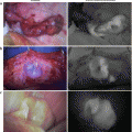
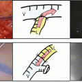
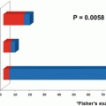
Fig. 6.1
Changes in OPCAB frequency rate of initial elective CABG in Japan
Since off-pump CABG is considered to be a more technically demanding surgery than traditional CABG, graft patency rate highly depends on the surgeon’s skill level. The method for the graft assessment was either intraoperative angiography using portable digital subtraction angiography (DSA) or postoperative catheter angiography, both of which involve X-ray radiation exposure for the patient and staff members, potantial renal injury to patients due to the use of iodinated contrast media, and other complications due to the need for percutaneus catheter insertion. Furthermore, even if postoperative angiography were to comfirm graft occlusion, it would be impossible to immediately reopen the patient’s chest to repair the technical graft failure. The SPY System (Novadaq Technologies Inc., Toronto, Canada) is the first commercially available fluorescence angiographic device which allows surgeons to assess the bypassed graft during the operation while the chest is still open without the concerns for radiation exposure or potentially harmful contrast agents. Intraoperative finding of technical errors using SPY system enables surgeons to revise the graft anastomosis during the operation.
6.2 About SPY System
The trend for the treatment of coronary artery diseases has been shifting from surgical CABG to percutaneous catheter intervention (PCI) with drug-eluting stents. The main reason for shifting to PCI may be its less invasiveness and immediate visual confirmation of the effectiveness of the treatment during its procedure through the use of X-ray angiography in the catheterization laboratory. As shown in Fig. 6.2, successful results can be confirmed quickly and easily during PCI; however, traditionally graft patency or blood flow cannot be seen without significant difficulty during CABG surgery. Moreover, angiographic confirmation of the results of PCI can be seen by the human eye, but anastomotic quality and blood flow through a graft cannot be seen by the surgeon’s eyes alone during CABG. In addition, there are other kinds of complementary technologies which can be used during PCI to evaluate the success of the procedure, such as intravascular ultrasound (IVUS), optimal coherence tomography (OCT), cardioangioscopy, and optical frequency domain imaging (OFDI) (Fig. 6.3).
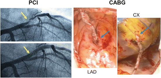
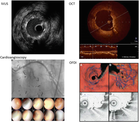

Fig. 6.2
Differences between PCI and CABG
Left: In PCI, blood flow and a successful result can be recognized at a glance using X-ray angiography.
Right: In CABG, using the human eye we can see there is no bleeding at the site of the graft anastomosis, but we cannot assess the quality of blood flow within the graft or graft patency

Fig. 6.3
Various imaging methods support procedure success in PCI
In order to make CABG a standard treatment option for the coronary artery diseases, it has been believed that highly useful intraoperative graft assessment tools are essential to sustain higher rates of graft patency. With the exception of X-ray angiography which is extremely difficult to use during CABG, there had never been reliable easy to use tools to visualize blood flow in the native coronary arteries and bypass graft particularly under the beating-heart OPCAB circumstance. The SPY system (Fig. 6.4) is an innovative intraoperative graft patency assessment device that is ideal for use during both traditional CABG and OPCAB because of its ease of use and reliability. The SPY system was first introduced to the Asia Pacific region and to this author in 2002 [4, 5]. Since that time, the system has been further developed to improve the quality of the image, the ease of image capture, to provide objective analysis tools and to add other features that meet the needs of surgery.
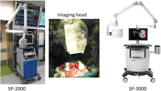

Fig. 6.4
SPY Intraoperative Imaging System
Left: First generation SPY system (SP-2000)
Center: In surgery, the imaging head is draped fixed 30 cm above the heart
Right: The latest generation SPY system (SP-3000)
The main characteristics of SPY system are:
1.
It enables surgeons to visually assess blood flow in vessels and graft patency immediately after the anastomosis is created.
2.
The real-time image of flow through the graft is equal or even superior to an X-ray angiogram.
3.
It does not involve the need for catheter insertion, ionizing radiation exposure or potentially nephrotoxic contrast media.
How to capture SPY image:
1.
After creating the anastomosis, the camera head is positioned approximately 30 cm above the area of the interest (Fig. 6.4 center).
2.
0.5 ~ 1.0 ml of 20 times diluted indocyanine green (ICG, 1.25 mg/ml) is injected through the central venous line followed by a 10 ml brisk bolus of saline(Fig. 6.5).
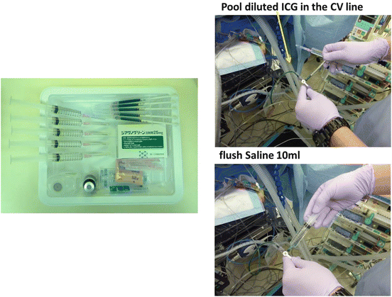

Fig. 6.5
How to inject the ICG imaging agent
Left: Prepare the required number of 0.5 ml or 1.0 ml of ICG diluted with 10 ml of aqueous solvent in 1.0 ml syringes and 10 ml of saline syringes for immediate bolus flush are prepared for SPY imaging
Right: Administer the diluted ICG in the central venous line, and flush it using 10 ml of saline
3.
10 ml of saline flush was administrated as a bolus injection following the ICG into the right atrium to ensure rapid circulation of the ICG. (Fig. 6.5).
4.
High quality fluorescence image will then be shown up on the monitor in a few seconds.
5.
ICG is excreted by the liver unchanged into the bile; hence, it does not have a negative affect on kidney function.
6.
SPY imaging is used not only to assess the graft and coronary arteries, it is possible to detect and verify the course of coronary veins of less than 0.5mm diameter.
7.
SPY imaging does not involve X-ray, but instead relies upon a laser (averaged output of 2.0 watt) to excite ICG which absorbs and emits light at 830nm which is then captured by the system’s CCD camera and the video output is displayed on the monitor.
The adverse drug reaction rate of ICG has been known to be much lower than the conventional iohexol iodine contrast media (Table 6.1), and it has been reported to be safely usable.
Table 6.1
Adverse drug reaction (ADR) reporting rate comparison between iodine contrast media and ICG
Contrast media (iohexol) | ICG | |
|---|---|---|
ADR reporting no./total no. | 452/17,039 | 36/21,278 |
ADR % | 2.65 % | 0.17 % |
Cardiovascular ADR % | 0.33 % | 0.023 % |
Image sequences of this ICG fluorescence imaging (Fig. 6.6) are captured and saved in the computer of the SPY system in either AVI or MPEG format. The latest version of SPY system allows saving the sequences in compressed DICOM format, which enables the users to export the data of more than 20 patients into one CD-R.
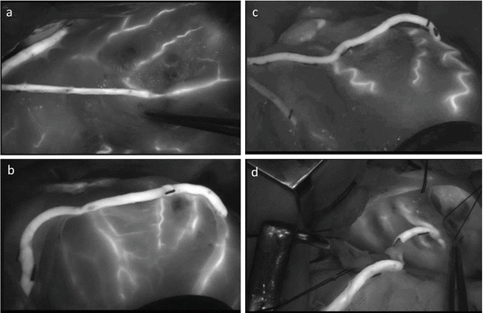

Fig. 6.6
Intraoperative SPY images
High quality graft images can be obtained wherever the distal anastomoses are performed in anterior, lateral, or inferior wall
(a) LITA-LAD, (b) SVG-HL-OM-PL, (c) RA-HL-OM, d GEA-4PD
6.3 Intraoperative SPY Images and Postoperative Angiography or MDCT
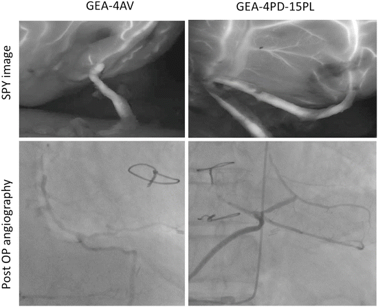
Several SPY system images utilizing the fluorescence properties of ICG were provided here (Figs. 6.7, 6.8, 6.9, and 6.10). The image quality of SPY system’s fluorescence angiogram including the internal thoracic artery (ITA), the sequential graft of radial artery (RA), saphenous vein graft (SVG), and gastroepiploic artery (GEA) is excellent. The quality of the SPY image appears equivalent to the conventional X-ray angiogram [6–10]. The actual real time moving SPY image sequences available on the SPY system monitor at the time of surgery are of even higher quality that those that can be published in this textbook. The blood flow inside the patet graft is highly visible. The head camera of the SPY system can rotate freely so that the circumflex (CX) site and the distal site of the right coronary artery (RCA) can be also visible. As shown in Fig. 6.11, similar images can be revealed between intraoperative SPY image and postoperative MDCT scanning.
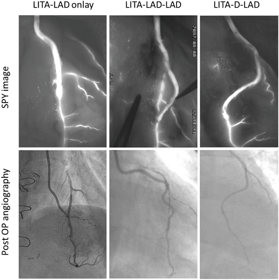
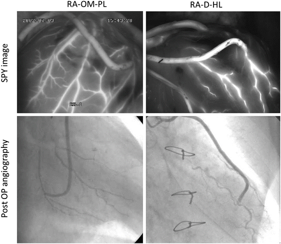
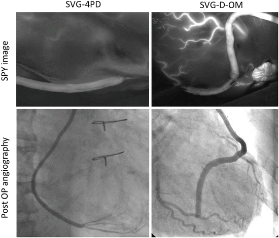

Fig. 6.7
SPY images and postoperative angiography of the left internal thoracic artery (LITA) graft
Almost same images could be seen in both SPY and angiography
Left: LITA-LAD on-lay patch graft
Middle: LTA-LAD-LAD sequential graft
Right: LITA-Diagonal-LAD sequential graft

Fig. 6.8
SPY images and postoperative angiography of the radial artery (RA) graft
Left: RA-OM-PL sequential graft
Right: RA-Diagonal-HL sequential graft

Fig. 6.9
SPY images and postoperative angiography of the saphenous vein graft (SVG)
Left: SVG-4PD
Right: SVG-Diagonal-OM sequential graft
