Fig. 23.1
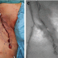
The macroscopic appearance of the mobilized upper esophagus, the whole stomach, and the ICG-AG findings
(a): The mobilized upper esophagus and whole stomach of the patient with long-gap congenital esophageal atresia. The only preserved feeding vessels for the mobilized whole stomach were the right gastric and right gastroepiploic arteries. The serosal surface color of both organs showed a normal color
(b) and (c): The ICG-AG findings obtained immediately following the injection of ICG. The good perfusion was confirmed with ICG-AG in the upper esophagus (b) and whole stomach (c), except for the small area in which the previous gastrostomy was closed (indicated with an arrowhead)
23.3.2 Case 2
A 15-year-old male underwent massive intestinal resection due to severe small intestinal volvulus. The residual intestine comprised only 37 cm of proximal jejunum and 7 cm of ileum. The serosal surface color of the distal part of the residual jejunum (DPRJ, the area encircled with the dotted line in Fig. 23.2a) initially showed a slightly darker hue than normal. After several cycles of irrigation of this segment with warm normal saline, the color of the DPRJ improved with time, and the other clinical findings also improved, which was considered to indicate that the perfusion of the DPRJ was preserved. The perfusion of that area was therefore clinically expected to improve with time.
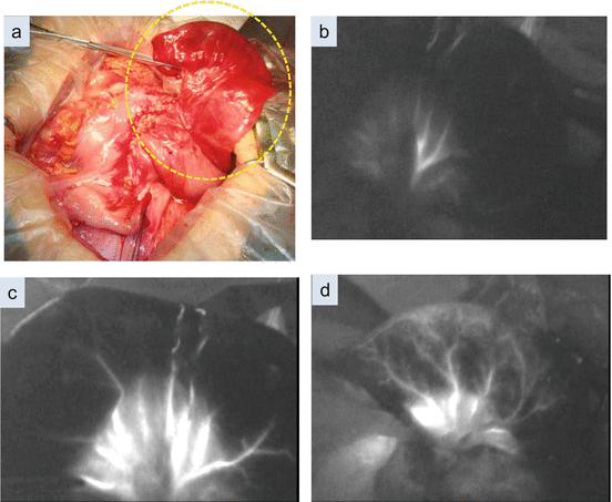

Fig. 23.2
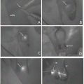
The macroscopic appearance of the residual jejunum following massive intestinal resection and the ICG-AG findings over time
(a): The residual jejunum and ileum immediately following massive necrotic intestinal resection. The serosal surface color of the distal part of the residual jejunum (DPRJ, the area encircled with the dotted line) showed a slightly darker hue than normal. We considered that the serosal color change shown in (a) was caused by transient venous congestion following the release of the intestinal volvulus and could be expected to improve over time
(b) and (c): These images correspond to the DPRJ in which the serosal surface exhibited a slightly darker hue (indicated with the encircled dotted line in (a)). Immediate, homogenous visualization of the mesentery was confirmed; however, the same findings were not observed in the intestine except for visualization of only a few main trunks of the vasa recta
(d) The findings of ICG-AG about 10 min after the initial intravenous ICG administration at the DPRJ. The capillary vessels communicating with the vasa recta were also gradually visualized
However, the first ICG-AG demonstrated immediate visualization of only a few main trunks of the vasa recta at the DPRJ, which exhibited a slightly darker hue on the serosal surface (Fig. 23.2b and c). Even after the apparent improvement of the color of the DPRJ was obtained, ICG-AG still showed only thin visualization of the vasa recta, including the leading capillary vessels (Fig. 23.2d). Homogeneous visualization of ICG fluorescence was not obtained during this examination. In contrast, in the proximal part of the residual jejunum, the immediate homogenous perfusion of ICG could be observed.
At this time, we finally estimated that the perfusion of the DPRJ was preserved, mainly based on the improvement of the clinical findings of the intestine in spite of the abnormal findings of ICG-AG. The primary anastomosis was performed without additional resection in order to maximize the lengths of the residual intestine. After the initial surgery, the patient developed a delayed partial stricture of the residual intestine, and an additional resection was performed on the 22nd postoperative day. The stricture segment corresponded to the area that presented abnormal findings by ICG-AG at the initial surgery, and this was resected at the second surgery.
The quantitative analysis of changes in the ICG fluorescence intensity over time that was performed on the day after the second surgery showed that the fluorescence intensity at the mesentery gradually increased over time, whereas that observed at the DPRJ demonstrated only a persistent slow increase in intensity and the absence of gradual increases over time (Fig. 23.3). We considered that the pattern of the changes in the ICG fluorescence intensity of the mesentery corresponded to the delayed drainage pattern described by Matsui et al. [6], whereas that of the DPRJ corresponded to the capillary or arterial insufficiency pattern.
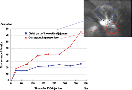

Fig. 23.3


The result of the retrospective quantitative analysis of the changes in the ICG fluorescence intensity at the DPRJ over time after ICG injection at the time of the initial surgery
The fluorescence intensity at the mesentery gradually increased over time, whereas that observed at the DPRJ demonstrated only persistent slow increases in intensity and the absence of gradual increases over time
Stay updated, free articles. Join our Telegram channel

Full access? Get Clinical Tree


