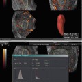
Fig. 12.1
Saline sonogram showing intrauterine adhesions
Management of IUA
Diagnosis and treatment of intrauterine adhesions are integral to the optimization of fertility outcomes [15]. Surgical management of IUA presents a challenge to the hysteroscopic surgeon. Though the appropriate management is controversial [3], and more often than not, guided by the clinician’s choice, skill, and operative setting, hysteroscopic adhesiolysis with antibiotic prophylaxis followed by the use of postoperative adjuvants such as systemic estrogens and intrauterine devices or systems designed to impede the development of adhesions is the treatment of choice with favorable results in terms of pregnancy and live birth rates [3, 15, 32, 33]. Clinicians should maintain a level of suspicion of intrauterine adhesions and should investigate by hysteroscopy if necessary [32]. Non-hysteroscopic techniques are also beginning to be developed, but whether they will replace the current “gold” standard of hysteroscopy remains to be seen [34]. The success of treatment regarding term deliveries and rate of abortions depends on the severity of the adhesions, and pregnancy, when achieved, may be complicated by premature labor, placenta previa, and placenta accreta [33].
Hysteroscopic Surgery
Technological progress in optic fibers and instrumentation has made it possible to video endoscope and determine the fibrous nature of the lesions and its precise localization and control endocavitary surgeries such as hysteroscopic adhesiolysis for uterine synechiae [35]. Though sonohysterography and hysterosalpingography are useful as screening tests of intrauterine adhesions [15], hysteroscopy has been considered the mainstay of diagnosis, classification, and treatment of the intrauterine adhesions, with medical treatments having no role in management [1, 2, 4, 15, 32, 36]. Diagnostic and therapeutic hysteroscopy is a simple, feasible, safe, reproducible, effective, quick, well-tolerated, and low-cost surgical procedure that is highly successful in an outpatient setting, offering a see-and-treat approach in majority of the subjects with intrauterine adhesions [37, 38]. Hysteroscopy has also become accepted as the optimum route of surgery, the aim being to restore the size and shape of the uterine cavity, normal endometrial function, and fertility [15, 16]. Lysis of intrauterine adhesions, for the treatment of infertility and recurrent pregnancy loss, results in improved fecundability and decreased pregnancy loss. Though adhesiolysis for pain relief appears efficacious in certain subsets of women, unfortunately, even when lysed, adhesions have a great propensity to reform [16]. According to Bettocchi et al. [38] there is no consensus on the effectiveness of hysteroscopic surgery in improving the prognosis of subfertile women. However, office hysteroscopy is a powerful tool for the diagnosis, and treatment of intrauterine benign pathologies and in patients with at least two failed cycles of assisted reproductive technology, diagnostic hysteroscopy and, if necessary, operative hysteroscopy is mandatory to improve reproductive outcome [38]. A descriptive study (Canadian Task Force classification II-2) concluded that hysteroscopic adhesiolysis is an effective and safe option even for postmenopausal women with intrauterine lesions adhesions on hysteroscopy or ultrasound. It allows the correct diagnosis to be made, reduces the need for major and unnecessary surgery, and is therapeutic in most patients [39].
Treatment can range from simple cervical dilatation in the case of cervical stenosis, but an intact uterine cavity, to extensive adhesiolysis of dense intrauterine adhesions using scissors, electro- or laser energy, or a combination of blunt and sharp dissection [32, 34]. Various techniques for adhesiolysis and for prevention of scar reformation have been advocated. According to March, [2] the use of miniature scissors for adhesiolysis and the placement of a balloon stent inside the uterus immediately after surgery appear to be the most efficacious [2]. Patients with more severe adhesions, in whom the uterine fundus is completely obscured, and those with a greatly narrowed fibrotic cavity present the greatest therapeutic challenge. Several techniques have been described for these difficult cases, but the outcome is far worse than in patients with mild, endometrial-type adhesions [4, 34]. A significantly obliterated cavity may require multiple hysteroscopic adhesiolysis to achieve a satisfactory anatomical and functional result [15, 36], while laparoscopic or ultrasound guidance may aid in the hysteroscopic lysis of dense scar tissue and difficult entry into the cervix [1].
Treatment Outcome
Treatment outcomes are difficult to assess as there is no universally agreed upon classification system [1]. Anatomic, but most of all functional prognosis, is directly correlated to the severity of adhesions, and the number of surgical procedures required to complete treatment [40].
Restoration of menstruation is highly successful (more than 90 %), and pregnancy rates around 50–60 % with live birth rates around 40–50 % can be achieved [32]. The risk of complications for those that achieve pregnancy is significant with a significant risk for placenta accreta and subsequent blood loss, transfusion, and hysterectomy [12]. In perhaps the largest study, involving 6,680 hysteroscopies with hysteroscopic adhesiolysis in 75 patients, 94.6 % functional restoration and 93.3 % anatomic resolution, with pregnancy rates ranging from 28.7 to 53.6 %, were achieved. At 2-month follow-up, the uterine cavity was completely regular in 70 cases, while in four cases, a second surgical treatment was necessary [41].
Using a standard technique with a loop electrode and glycine 1.5 % as distension medium, Dawood et al. [12] reported an improvement in the rate of amenorrhea from 32.3 % before adhesiolysis to 9.2 % after the procedure with an overall pregnancy rate of 51.2 % and the live birth rate 32.6 % among women who wished to conceive. Severe intrauterine adhesions were managed with the assistance of abdominal ultrasound to ensure that the uterine cavity was not breached, and the rates of pregnancy and term pregnancy among this selected group of women were similar regardless of the severity of adhesions [12].
Yu et al. [42] evaluated the outcome of hysteroscopic adhesiolysis with electrode needle or loop under direct vision in 85 women with Asherman’s syndrome who presented with a history of infertility or recurrent pregnancy loss. After hysteroscopic adhesiolysis, the chances of conception among the 18.2 % of women who remained amenorrheic were significantly lower than those who continued to have menses (50 %). The conception rate in women who had reformation of intrauterine adhesions at second look hysteroscopy (11.8 %) was significantly lower than that of women who had a normal cavity (59.1 %), suggesting that the outcome of hysteroscopic adhesiolysis for Asherman’s syndrome is significantly affected by recurrence of intrauterine adhesions [42].
Hysteroscopic adhesiolysis with monopolar or bipolar energy can be performed safely and effectively for severe stage 3 and 4 adhesions with a 97 % restoration of menses, 43.8 % PR, and 32.8 % LBR. The pregnancy rate was significantly higher in patients ≤35 years compared to patients older than 35 years (66.6 % vs. 23.5 %, respectively; p = 0.01), suggesting that age is the main predictive factor of success: the pregnancies were at risk of abnormal placentation [43]. The impact of age on the outcome of hysteroscopic adhesiolysis is in agreement with a previous study by Capella-Allouc et al. [44] that reported a pregnancy rate of 42.8 %, live birth rate of 32.1 %, the pregnancy rate being much higher in patients ≤35 years compared to patients older than 35 years (62.5 % vs.16.6 %, respectively; p = 0.01) following hysteroscopic adhesiolysis in 31 patients with severe Asherman’s syndrome. However, these pregnancies were at risk for hemorrhage with abnormal placentation [44].
Roy et al. [10] reported an overall conception rate of 40.4 %, live birth rate of 86.1 % and a miscarriage rate of 11.1 % in a mean conception time after surgery of 12.8 months following hysteroscopic adhesiolysis with the monopolar electrode knife in 89 infertile patients with Asherman’s syndrome. The cumulative pregnancy rate showed that 97.2 % of patients conceived within 24 months. The conception rate was higher (58 %) in mild Asherman’s syndrome compared to 30 % conception rate in moderate and 33.3 % conception rate in severe cases. There was a significantly higher likelihood of conception (44.3 %) in those who continued to have improved menstrual pattern compared to only 10 % likelihood of conception in those who continued to have amenorrhea after adhesiolysis. A second-look office hysteroscopy, performed after 2 months, showed reformation of adhesions in12 patients that needed a repeat adhesiolysis with no conception in these patients. The authors concluded that hysteroscopic adhesiolysis for Asherman’s syndrome is a safe and effective method of choice for restoring menstrual function and fertility [10].
Shokeir et al. [45] attempted to analyze the adhesion grade in multiple hysteroscopic-guided biopsies from IUA following the initial hysteroscopic adhesiolysis at a follow-up diagnostic hysteroscopy, performed early (2–4 weeks) after the initial operation or late, about 12 months (8–16 months). They observed that at follow-up hysteroscopy, 25 % of both groups had no significant adhesions. Grade I adhesions (thin, filmy) occurred in 60 % of the early hysteroscopy patients and in only 12 % of the late group (P < 0.05). Grade II adhesions were present in 10 % of the early group and in up to 41 % in the late group (P < 0.05), whereas grade III adhesions were present in only 5 % of the early hysteroscopy group, but in 22 % of the late one (P < 0.05). Correlation between hysteroscopic and histologic findings were good in most of cases in both groups. The follow-up to determine the subsequent reproductive outcome revealed similar conception rates in both groups. The authors suggested that the IUA that might be formed immediately following hysteroscopic reproductive surgery is histologically different from those appearing a longer time after the original operation. Routine early follow-up hysteroscopy can influence the prognosis resulting from the original surgery [45].
Having excluded hormonal imbalances, premature ovarian failure, and congenital uterine abnormalities Yasmin et al. [46] reported thick fibrous adhesions in 45 % of patients, flimsy adhesions in 40 %, and muscular adhesions in 15 % at hysteroscopy, with 65 % adhesions in the body of uterus, 25 % at the site of internal os, and 1 % had adhesions in the cervical canal as well as the body of the uterus. Following diagnostic hysteroscopy and resection of adhesions in 20 patients (median age 26 years), presenting with scanty menses and secondary infertility (65 %), secondary amenorrhoea (20 %), or with primary infertility alone (15 %), they reported a restoration of menses in 95 % of the patients and conception in 10 % of the patients. Though the patient number was small, the authors suggested that hysteroscopy is an effective procedure for not only diagnosing Asherman’s syndrome, but is equally effective for treating it [46].
Hysteroscopic adhesiolysis in women with Asherman’s syndrome and poor reproductive performance (previous spontaneous abortions or a premature delivery) contributes significantly to a successful reproductive outcome. Whereas pregnancy outcome prior to the hysteroscopic adhesiolysis was 18.3 % term deliveries, 3.3 % premature deliveries, 62.4 % first-trimester abortions, and 16.0 % late abortions, after hysteroscopic adhesiolysis, the pregnancy outcome was 68.6 % term deliveries, 9.3 % premature deliveries, 17.4 % first-trimester abortions, and 4.7 % late abortions. The operative success rate, measured by delivering a healthy newborn, improved from 18.3 % preoperatively to 64 % postoperatively in women with two previous unsuccessful pregnancies [47], whereas in women with three or more unsuccessful pregnancies, the success rate improved from 18.3 to 75 %. Successful outcome of adhesiolysis was observed in 61.9 % of mild (stage I) and in 70.6 % of moderate to severe cases (stages II and III) of intrauterine adhesions [47].
Blunt adhesiolysis with a flexible hysteroscope, following primary treatment of intrauterine adhesions with sharp adhesiolysis has been suggested as an effective technique for the maintenance of cavity patency with an improvement in menstrual flow in 95 % of the patients, relief of dysmenorrhea in 92 %, 92 % improvement in disease staging over the treatment interval, and a pregnancy rate of 46 %. Initially, 50 % had severe adhesions, 46 % had moderate, and 4 % had minimal disease according to the March criteria [48].
Colacurci et al. [49] analyzed the reproductive outcome in 53 women undergoing hysteroscopic lysis of intrauterine adhesions, according to their localization and severity. Hysteroscopic surgery restored an acceptable menstrual cycle in almost all the patients affected by intrauterine isolated adhesions in 52 % of women with complex incomplete adhesions, and in none of the patients with an entirely obliterated cavity. In isolated, isthmic, central, or marginal synechiae, a pregnancy rate of 73.3 % was observed with a pregnancy rate to term, respectively, of 63.3 % and of 86.3 %, while in case of complex but not complete adhesions, the pregnancy rate was 25 % with only two term pregnancies. There were no pregnancies in three cases of complex synechiae. The authors concluded that the basic parameter to define the functional and reproductive prognosis of the hysteroscopic lysis of intrauterine adhesions is not the menstrual profile or the histological characteristic of the lesions, but rather their extension [49].
Hysteroscopy and hysteroscopic surgery have been the gold standard of diagnosis and treatment, respectively, for patients with Asherman’s syndrome who presented with amenorrhea or hypomenorrhea, infertility, or recurrent pregnancy loss. However, according to most authors, despite the advances in hysteroscopic surgery, the treatment of moderate to severe Asherman’s syndrome still presents a challenge [40, 50]. Furthermore, pregnancy after treatment remains high risk with complications including spontaneous abortion, preterm delivery, intrauterine growth restriction, placenta accreta or previa, or even uterine rupture, that necessitate close antenatal surveillance and monitoring for women who conceive after treatment [50]. According to Piketty et al. [40] despite the infrequent but well-known complications during surgery and the less frequent but often severe obstetrical complications, the benefit gained by the recovery of fertility (either spontaneous or not) remains superior to the risks of the surgical management [40].
Role of Ultrasonography in the Treatment
Serial intrauterine device-guided hysteroscopic adhesiolysis of intrauterine synechiae, especially for early intervention, may prevent complications during the treatment of severe intrauterine adhesions and may present a secure and effective alternative for constructive clinical outcomes with spontaneous pregnancy rates of 47.2 and 30 % and live birth rates of 28 and 20 % in patients who did and did not undergo early intervention of office hysteroscopy, 1 week after insertion of the IUD at hysteroscopic adhesiolysis, respectively [51]. Following echo-controlled hysteroscopic surgical cure of complex and/or recurrent uterine synechiae in 11 patients, Salat-Baroux et al. [35] concluded that intraoperative echography allowed hysteroscopic adhesiolysis of intrauterine adhesions at a controlled and equivalent distance from the uterine walls, enabling better treatment of the uterine cornua since the operator is informed when to limit progression to avoid massive fluid infusion into the abdominal cavity and perforation of the uterus. The intraoperative echographic control was validated in the operating theater radiographically. With this technique normal cavities with bilateral tube permeability were obtained in 72.72 % of the patients and normal cycles in 90.9 % of the patients [35]. Following hysteroscopic lysis under ultrasound control for significant intrauterine synechiae, Bellingham [52] reported normal menstruation in 61 % of the patients and live births in 80 % of the patients, of whom 50 % had had severe adhesions. They reported that ultrasound control is ideally essential if the adhesions are extensive [52]. However, in both these studies, the number of patients was very small to effectively document the role of ultrasound in the treatment of IUA.
Coccia et al. [53] described a new therapeutic procedure called pressure lavage under ultrasound guidance (PLUG) for selected cases of IUA. This technique is based on sonohysterography to monitor the effects of intrauterine injections of saline solution on the continuous accumulation of saline in the uterine cavity for the mechanical disruption of IUA. In an open clinical investigation with no control group, they reported satisfactory lysis of adhesions and restoration of menses in 71.4 % of the patients with mild IUA with a pregnancy rate of 66.63 % following the use of the PLUG technique. A second-look hysteroscopy after 1 month showed the persistence of filmy adhesions in two patients with moderate IUA that were removed successfully during hysteroscopy. The authors suggested that PLUG is a safe and ideal in-office procedure that allows complete lysis in mild IUA cases avoiding the need for therapeutic, and possibly, follow-up hysteroscopy, and may represent a useful initial step in moderate IUA cases reducing the need for operative hysteroscopy [53]. In a recent study, Taniguchi and Suginami [54] also suggested that sonohysterographic (SHG) lysis for recurrent adhesions following hysteroscopic lysis may be a treatment option for recurrent adhesions in infertile patients, with improved menstrual cycles and restored tubal patency [54].
Tiras et al. [55] demonstrated the value of laparoscopic intracorporeal ultrasound (LIU)-guided hysteroscopic adhesiolysis in a patient with amenorrhea and infertility with total intrauterine synechiae. Adequate intrauterine adhesiolysis was performed by a resectoscope with a wire loop, suggesting that complex intrauterine procedures can be easily performed by the guidance of endoscopic ultrasonography to avoid the possibility of inadvertent uterine perforation [55].
Schlaff and Hurst [56] evaluated the predictive value of preoperative endometrial sonography in the diagnosis and surgical treatment of women with amenorrhea due to severe Asherman’s syndrome, characterized by complete obstruction of the cavity at hysterosalpingogram. They suggested that an endometrial pattern, demonstrating a well-developed endometrial stripe on transvaginal sonography is highly predictive of a positive surgical and clinical outcome in women with severe Asherman’s syndrome with resumption of normal menses and normalization of the cavity after hysteroscopy in contrast to women with minimal endometrium who had no cavity identified and derived no benefit from surgery [56]. However, this study was limited to just seven patients and hence, substantial evidence in this direction is lacking.
Radiographic Methods
In a small but significant study, Karande et al. [57] demonstrated that in-office lysis of intrauterine adhesions, under fluoroscopic control, using a specially designed catheter (gynecoradiologic control) can be carried out safely in the majority of patients, using minimally invasive techniques. They could successfully lyse adhesions in 76 % (13/17) of the patients (9 mild, 3 moderate, and 1 severe), while in remaining 4 patients (2 moderate and 2 severe), lysis was only partially successful. Nine procedures were performed with the catheter’s balloon tip and four with hysteroscopic scissors. Procedure complications resulting in the abandoning of the procedure included patient discomfort before attempting the use of scissors (n = 1), extravasation of dye into the myometrium making visualization difficult (n = 1), and thick, fibrotic adhesions that were resistant to scissors (n = 2). They opined that the potential cost savings with this technique in comparison with endoscopic procedures, which require utilization of expensive operating room time, are especially relevant in a cost-conscious managed care environment and only failures of in-office procedures would reach the operating room. [57] The fluoroscopic approach to adhesions was further evaluated a decade later by Chason et al. [58] who used hysteroplasty with fluoroscopic cannulation and balloon uterine dilation to treat intrauterine adhesions and cervical stenosis and lower uterine defects in select cases. They concluded that while the treatment of intrauterine adhesions resulted in an improved pregnancy outcome, albeit in a case study, the effect of lower uterine segment-filling defects from cesarean deliveries on pregnancy outcome in assisted reproductive technology cycles warrants further investigation [58]. In a 5-year retrospective, uncontrolled cohort study, Thomson et al. [59
 Two-Dimensional and Three-Dimensional Doppler in Reproductive Medicine
 Ultrasound and PCOS
 Focused Ultrasound for Treatment of Fibroids
 Ovarian Reserve and Ovarian Cysts
 Virtual Hysterosalpingography: A New Diagnostic Technique for the Study of the Female Reproductive Tract
 The Normal Ovary (Changes in the Menstrual Cycle)


Related posts:
Two-Dimensional and Three-Dimensional Doppler in Reproductive Medicine
Ultrasound and PCOS
Focused Ultrasound for Treatment of Fibroids
Ovarian Reserve and Ovarian Cysts
Virtual Hysterosalpingography: A New Diagnostic Technique for the Study of the Female Reproductive Tract
The Normal Ovary (Changes in the Menstrual Cycle)
Stay updated, free articles. Join our Telegram channel
Full access? Get Clinical Tree
