Fig. 1.1
The joints of the wrist: the distal radioulnar (a) the radiocarpal (b) and the mediocarpal (c) joints. There is normally no communication between the three joints
The radioulnar joint is also involved in pronosupination of the wrist.
Of the various structures composing the wrist (bones, synovial membranes, joint capsules, etc.), the tendons are the ones most frequently examined with ultrasonography. They can be divided into two main groups: dorsal and volar. The tendons on the dorsal aspect of the wrist (Table 1.1) run through 6 osseofibrous tunnels [3–5] (Fig. 1.2) or compartments. The floor of these tunnels is formed by the radius, the ulna, and the distal radioulnar joint; the roof consists of the extensor retinaculum (also known as the dorsal carpal ligament).
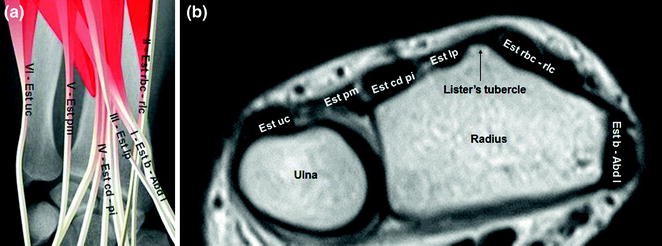
Table 1.1
Osseofibrous tunnels (or compartments) on the dorsal aspect of the wrist
Compartment 1 | Abductor pollicis longus and extensor pollicis brevis tendons |
Compartment 2 | Extensor carpi radialis longus and extensor carpi radialis brevis tendons |
Compartment 3 | Extensor pollicis longus tendon |
Compartment 4 | Extensor digitorum communis and extensor indicis proprius tendons |
Compartment 5 | Extensor digiti minimi proprius tendon |
Compartment 6 | Extensor carpi ulnaris tendons |
Fig. 1.2
Tendons of the dorsal wrist: a schematic diagram; b axial T1-weighted MRI scan shows (moving from the radial to ulnar sides): the extensor pollicis brevis and abductor pollicis longus (Est b—Abd l); extensor carpi radialis brevis and extensor carpi radialis longus (Est rbc—rlc); the extensor pollicis longus (Est lp); the extensor digitorum communis and extensor indicis proprius (Est cd pi); the extensor digiti minimi proprius (Est pm); and the extensor carpi ulnaris (Est uc)
Each compartment-regardless of the number of tendons it contains is lined with a single synovial sheath . On the volar side of the wrist, there are four muscle layers with their respective tendons [3, 4] (Table 1.2) (Fig. 1.3). The most superficial layer includes the flexor carpi radialis, the flexor palmaris longus, and the flexor carpi ulnaris (Fig. 1.3a); the second layer consists of the flexor digitorum superficialis (Fig. 1.3b); the third includes the flexor digitorum profundus and flexor pollicis longus (Fig. 1.3c), and the fourth is composed of the pronator quadratus (Fig. 1.3d).
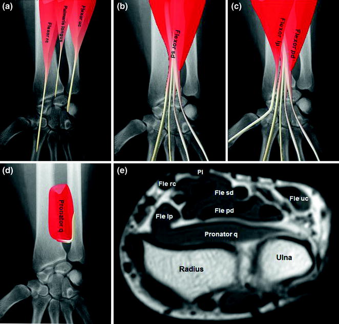
Table 1.2
Musculotendinous layers on the volar aspect of the wrist
Layer I | Flexor carpi radialis, palmaris longus, flexor carpi ulnaris |
Layer II | Flexor digitorum superficialis |
Layer III | Flexor digitorum profundus, flexor pollicis longus |
Layer IV | Pronator quadratus |
Fig. 1.3
Tendons of the volar wrist: a–d schematic diagrams; e axial T1-weighted MRI scan shows (from the radial to the ulnar side of the wrist) the superficial layer containing the flexor carpi radialis (Fle rc), palmaris longus (Pl), and flexor carpi ulnaris (Fle uc) (a, e); the second layer containing the flexor digitorum superficialis (Fle sd) (b, e); the third layer with the flexor digitorum profundus (Fle pd) and flexor pollicis longus (Fle lp) (c, e); and the fourth layer with the pronator quadratus (Pronator q) (d, e)
The tendons of the flexor digitorum superficialis and profundus muscles (four tendons each), the flexor pollicis longus tendon (which has its own synovial sheath), and the median nerve all lie within the carpal tunnel (Table 1.3), which is formed by the carpal bones and the flexor retinaculum (also known as the transverse carpal ligament) [2, 3] (Fig. 1.4); the flexor carpi radialis, flexor carpi ulnaris, and palmaris longus tendons lie outside this tunnel.
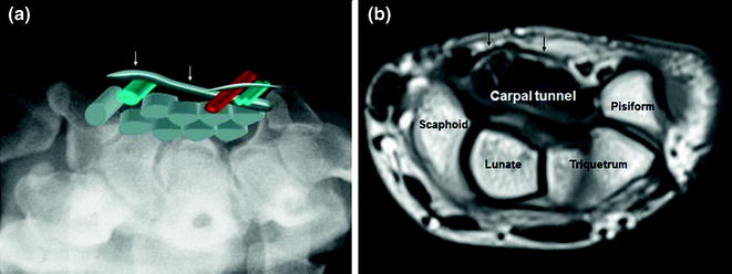
Table 1.3
Components of the carpal tunnel
• Carpal bones |
• Flexor retinaculum |
• Flexor pollicis longus |
• Flexor digitorum superficialis |
• Flexor digitorum profundus |
• Median nerve |
Fig. 1.4
The carpal tunnel: a schematic diagram; b axial T1-weighted MRI scan. The lateral and ventral walls of the carpal tunnel are formed by the bones of the wrist, and the flexor retinaculum (arrows) delimits it superficially. Inside the tunnel, the superficial and deep flexor tendons of the fingers (four of each) are enclosed within a single synovial sheath; they are accompanied by the flexor pollicis longus tendon enveloped in a sheath of its own; and the median nerve, which usually lies superficial to the flexor pollicis longus and to the 2 superficial flexor laminae for the index and middle fingers. The Guyon’s canal lies on the ulnar side, superficial to the carpal tunnel
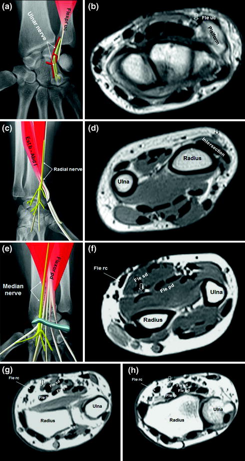
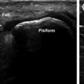
Superficial to the carpal tunnel lies the Guyon’s canal, a space formed by the flexor retinaculum (floor) and the palmar carpal ligament (roof). The ulnar artery and ulnar nerve both pass through this canal (Fig. 1.4a).
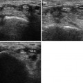
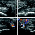
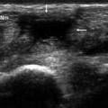
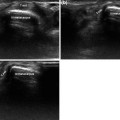
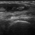
In addition to the ulnar nerve (Figs. 1.4a, 1.5a and b), the wrist also contains the radial (Fig. 1.5c and d) and median nerves (Fig. 1.5e–h).
Get Clinical Tree app for offline access
Fig. 1.5
Nerves of the wrist: the ulnar (a, b), radial (c, d), and median (e–h) nerves. The ulnar nerve (open arrow) runs through the Guyon’s canal, usually between the pisiform (ulnar side) and the ulnar artery (radial side) (a). At this level, it has already divided into a superficial, sensory branch and a deep, motor branch. The radial nerve (open arrow) (c, d) runs deep in the forearm, in close contact with the radius. In the wrist, it becomes more superficial and gives rise to its terminal sensory branches after crossing over the myotendinous junctions of the abductor longus and the extensor brevis. At this point, the latter tendons pass over the long and short radial extensors of the wrist (c). The median nerve (open arrow


Related posts:
Stay updated, free articles. Join our Telegram channel
Full access? Get Clinical Tree
