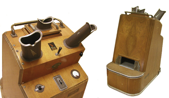
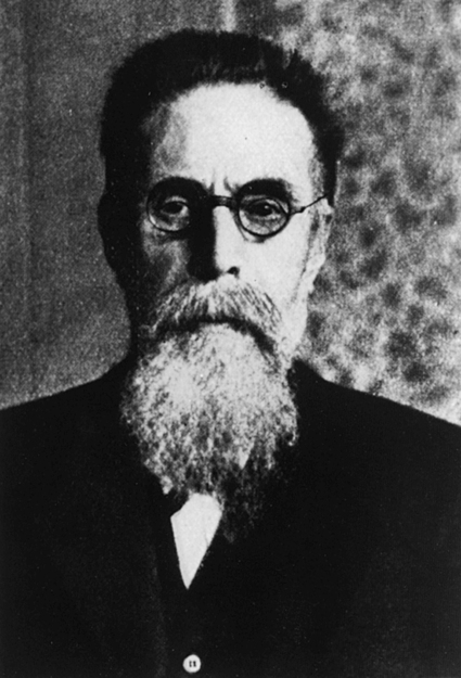
• Discuss key events in the discovery and evolution of the use of x-rays. • Apply general physics fundamentals, including recognition of units of measure and basic calculations. • Define and use radiologic units of measure. • Identify the general components of permanently installed radiographic equipment. • Describe the basic role and function of the general components of a permanently installed radiographic unit. Dr. Wilhelm Conrad Roentgen (Figure 1-1) was born March 27, 1845, in Lennep, Germany. His public education and academic career were marked by struggle, not for lack of intelligence but for want of opportunity. Following an unfortunate prank perpetrated by a classmate, he was expelled from school because he would not name the perpetrator. This began his struggle to find a place in a university to study. He eventually triumphed, receiving his PhD degree from the University of Zurich in 1869. He did, however, continue to struggle initially to establish himself as a professor and academician. Again, as a credit to his scientific skill and knowledge, he achieved considerable success, most notably being named director of the then newly formed Physics Institute at the University of Wurzburg in 1894. It was in this “state of the art” laboratory (for its time) that Dr. Roentgen forever changed the world of medicine. Late on a Friday afternoon, November 8, 1895, Dr. Roentgen was working in his laboratory. He had prepared a series of experiments involving a cathode ray tube of the Crookes’s type (it may have been a Hittorf tube, but the general design and features of both types are the same). The nature of cathode rays was of interest to many scientists of the day, and much experimentation was being conducted. On this particular evening, after setting up the tube and preparing for the evening’s experiments, Dr. Roentgen completely covered the tube with black cardboard to continue his study of the fluorescent properties of the cathode rays. On a table a few feet away was a piece of cardboard painted with barium platinocyanide. On beginning his experiments, he noticed that the piece of cardboard fluoresced each time the tube was energized. He had already verified that the cause could not be the visible light because he had covered the tube with the black cardboard and checked to be sure no light escaped. He also knew, according to the common knowledge of the day, that the cathode rays could not penetrate the glass walls of the tube. He moved the barium platinocyanide–coated cardboard closer and started his fevered investigation of this unknown light. He was consumed by a desire to understand this phenomenon and spent the next 7 weeks investigating it. It is said that he even took his meals in his laboratory and had his bed moved there to facilitate his research. So thorough was his investigation that he described practically every property of x-rays that we know today. As a part of his investigation, he asked his wife to allow him to “photograph” her hand with this new x-light, and, on December 22, 1895, he produced the first radiograph (Figure 1-2), and a profession was born. In the early days, the cathode ray tubes and generators used for such exposures were inefficient and the x-ray output varied considerably in quantity and quality. Exposure times were commonly in the 20- to 30-minute range; some exposures took up to 2 hours. Because of this, the early ventures into medical imaging came at a price. Many patients and operators suffered from acute radiodermatitis (radiation burns). There were even cases of electrocution of the operator in setting up the equipment for exposure because the equipment was not enclosed and shielded as it is today (Figure 1-3). Thomas Edison brought some attention to the dangers of x-rays. He suffered a radiation burn to his face and injury to his left eye from his experimentation with x-rays and discontinued his investigations. Edison’s assistant, Clarence Dally, did not cease investigation and truly suffered for it. Because of his experiments, Dally developed severe radiation burns. The only treatment of his day for such injuries was amputation, and during the course of his experiments (1897–1903) his left hand above the wrist, four fingers of his right hand, his left arm above the elbow, and finally the right arm at the shoulder were all amputated. At the end of his life, he was in such pain that he could not lie down and in 1904 died an agonizing death. Many of the early injuries were to “technicians” (as they were initially called) and doctors who worked with x-rays, and amputations and gloved hands became an identifying trait of their profession (Figure 1-4). By 1900, improved imaging plates, equipment, and techniques had all but eliminated acute radiodermatitis, but there was still a rather carefree attitude toward investigation and use of x-rays. Within the medical community, recognition of the problems and early efforts to minimize them were underway, but x-rays had also captured the public’s imagination in other ways. Immediately following the discovery and announcement, the public imagination went wild with speculation. Imagine a ray that could see through human flesh! Hopes abounded for this new, mysterious light, and there was speculation that it would soon be incorporated into a machine that could miraculously cure a host of mortal ills. The term x-ray appeared as the subject of poems, songs, and plays. It also appears in advertisements for polishes, ointments, batteries, and powders, and the list goes on. Opportunistic advertisers and manufacturers took advantage of the glamour and mania of the word x-ray and incorporated it into a host of products. Advertisements claimed that “x-ray stove polish” would clean your stove better, “x-ray headache tablets” would cure your headache quickly, “x-ray prophylactics” would prevent a long list of diseases, and even “x-ray golf balls” would fly farther and straighter! Examples of such advertisements are presented in Figure 1-5. Of course x-rays had nothing to do with any of these products’ effectiveness, only their improved sales. There were, however, actual applications of x-ray machines. One such application was the shoe fitter (Figure 1-6). This was a fluoroscope apparatus placed in shoe stores to help with the proper fitting of shoes. The advertisement claimed that such machines were vital to ensure comfort through the proper alignment of the bones of the foot within the shoe. Some advertisements stated that this was of particular importance in fitting children’s shoes. Consider the radiation dose that a child might have received during such a fitting or while playing with the machine as entertainment while a mother and father shopped!
Introduction to the Imaging Sciences
Discovery and Use of X-Rays
Dr. Roentgen’s Discovery
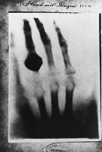
Image is of Dr. Roentgen’s wife’s hand. Note the ring on her fourth digit. (From Glasser O: Wilhelm Conrad Roentgen and the early history of the roentgen rays, 1933.)
Overview of X-ray Evolution and Use
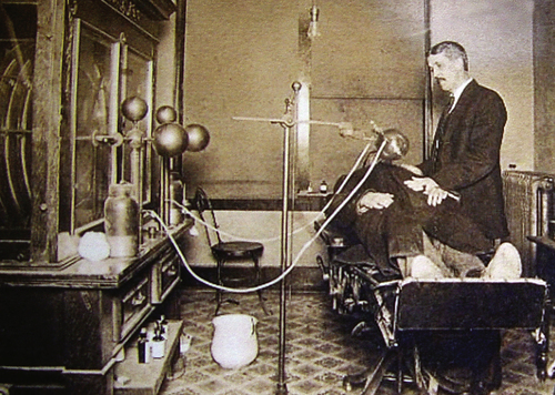
Image of circa 1900 x-ray machine setup in a physician’s office. Note the x-ray tube suspended above the patient and the open nature of the electrical wiring and tube. (Photo courtesy Alex Peck Medical Antiques.)
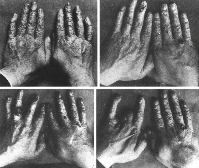
Picture of x-ray dermatitis and resultant amputations. Often gloves were worn to cover these injuries and amputations. (From Pancoast HK: Amer Quart Roentgen 1:67, 1906.)


Advertisements using the glamour of the word x-ray circa 1900.
Rights were not granted to include this content in electronic media. Please refer to the printed book. (Reprinted with permission from RadioGraphics and Dr. Ed Gerson.)
Introduction to the Imaging Sciences
 Critical Concept 1-1