Fig. 30.1
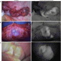
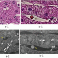
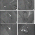
Three types of hepatocellular carcinoma (HCC) as determined by intraoperative laparoscopic indocyanine green (ICG) fluorescence imaging. (a) Laparoscopic view using standard light. (b) Whole positive fluorescence in the cancerous region. (c) The incised lesion also showed whole positive fluorescence in the cancerous region, which was diagnosed as HCC. (d) Laparoscopic view using standard light. (e) Partial positive fluorescence in the cancerous region. (f) The incised lesion also showed partial positive fluorescence in the cancerous region, which was diagnosed as HCC. (g) Laparoscopic view using standard light. (h) Ring positive fluorescence in the cancerous region. (i) The incised lesion also showed ring positive fluorescence in the cancerous region, which was diagnosed as HCC
In recurrent HCC, detection of the tumor margins, whether by visual inspection, palpation, or intraoperative ultrasound, is challenging in patients previously treated with transarterial chemoembolization (TACE) or radiofrequency ablation. Using ICG fluorescence imaging, viable cancer could be clearly distinguished from necrotic liver tissue following TACE (Fig. 30.2).
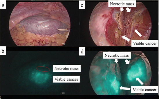

Fig. 30.2
Detection of the tumor margin in recurrent hepatocellular carcinoma (HCC) using laparoscopic indocyanine green (ICG) fluorescence imaging. (a) Laparoscopic view using standard light. (b) ICG-positive and ICG-negative fluorescence on the surface of the liver. (c) The area of positive fluorescence was diagnosed as HCC and the area of negative fluorescence as necrosis. (d) ICG-positive and ICG-negative fluorescence of the incised lesion
30.3.2 Intrahepatic Cholangiocellular Carcinoma
ICG fluorescence imaging of the liver surface clearly identified regions of cholestasis caused by tumor invasion of the bile duct or by thrombi.
In open laparotomy, fluorescence images of the liver surface were obtained intraoperatively with a fluorescence imaging system (PDE; Hamamatsu Photonics, Hamamatsu, Japan) composed of a charge-coupled device camera that excluded light with wavelengths of less than 820 nm and 36 light-emitting diodes with a wavelength of 760 nm [1, 4]. The cholestatic regions on the surface of the liver were clearly delineated with fluorescence imaging (Fig. 30.3), whereas using a laparoscopic system fluorescence in the same regions was relatively weak (Fig. 30.4). The latter result reflects the fact that, in the laparoscopic system, the fluorescence strength cannot be adjusted.
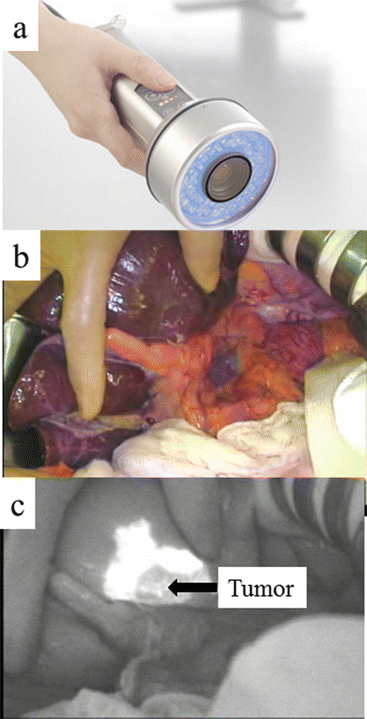
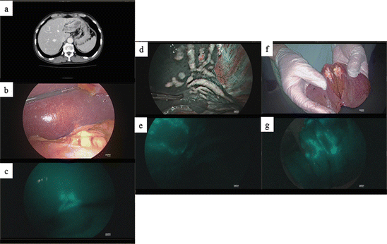

Fig. 30.3
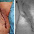
Fluorescence imaging of intrahepatic cholangiocellular carcinoma (ICC). (a) The fluorescence imaging system (PDE; Hamamatsu Photonics, Hamamatsu, Shizuoka) consisted of a charge-coupled device camera that filtered out light with wavelengths less than 820 nm and 36 light-emitting diodes with a wavelength of 760 nm. (b) Operative view using a standard light. (c) Intraoperative fluorescence imaging of cholestatic regions in ICC

Fig. 30.4


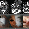
Laparoscopic fluorescence imaging of intrahepatic cholangiocellular carcinoma (ICC). (a) Preoperative contrast-enhanced computed tomography in a patient with ICC and a dilated bile duct in the left liver. (b) Laparoscopic view using standard light. (c) Ring positive indocyanine green (ICG) fluorescence in the cancerous region. (d) Laparoscopic view using standard light shows the dilated bile duct in the left liver. (e) Fluorescence imaging of the dilated bile duct. (f) The incised lesion as seen using standard light. (g) The incised lesion also showed ring positive fluorescence in the cancerous region, which was diagnosed as ICC
Stay updated, free articles. Join our Telegram channel

Full access? Get Clinical Tree


