Fig. 1
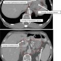
CT anatomy of the larynx. (a) Sagittal CT of the larynx. The pre-epiglottic space is anterior to the epiglottis and superior to the vocal folds. It is contiguous with the paraglottic space and base of tongue. The hyoid bone divides the epiglottis into infra- and suprahyoid portions. (b) Coronal CT demonstrates the false cord, the true cord, and the ventricle in between. (c) The paraglottic space is a small fat plane adjacent to the thryoid cartilage. (c, d) FVCs can be distinguished from the TVCs because the FVCs have a band of fatty tissue and the TVCs do not
The paraglottic and pre-epiglottic spaces are connected fat planes. The paraglottic space is bounded by the thyroid cartilage laterally and the TVCs, FVCs medially. The pre-epiglottic fat space is bounded by the mucosal surface of the vallecula superiorly, hyoid/thyroid strap muscles anteriorly, and root of the epiglottis posteriorly and inferiorly communicates with the paraglottic spaces.
Laryngeal cancer often invades the paraglottic and pre-epiglottic spaces. There are no barriers to spread to the adjacent space if one is involved.
The AJCC 6th edition added paraglottic space invasion to the staging system, denoting laryngeal tumors with this clinical feature as T3. Some practitioners continue to treat a T3N0 glottic SCC (T3 due only to paraglottic invasion) with limited fields (Dagan et al. 2007).
The thyroid cartilage has an inner and outer cortex. Invasion of the inner cortex only signifies T3 disease and invasion through the outer cortex signifies T4 disease. The degree of invasion can only be assessed through CT imaging with appropriate windowing and must be carefully reviewed by the treating radiation oncologist.
Surgeries relevant to advanced laryngeal cancer include total laryngectomy, supraglottic laryngectomy (also called horizontal partial laryngectomy), and vertical hemilaryngectomy transoral laser microsurgery (TLM). Supraglottic laryngectomy is appropriate only for cases without vocal fold fixation, involvement of the arytenoids, and/or thyroid cartilage invasion. Patient must also have good respiratory function in case of aspiration postsurgery. Historically, the inferior extent of the tumor must not exceed the level of the FVCs for this surgery, as the inferior surgical margin is the ventricles.
In general TLM is contraindicated for T3 or T4 disease.
2 Diagnostic Workup Relevant for Target Volume Delineation
Critical parts of the history include an assessment of voice, swallowing (i.e., modified barium swallow), respiratory, and constitutional status. A history of an emergent tracheostomy for respiratory dysfunction is a recurrence risk factor for T3 cases using organ preserving radiation, and thus total laryngectomy is a reasonable recommendation. Smoking cessation interventions should be instituted.
On examination, the larynx should be palpated and gently moved. Very advanced disease may exhibit absence of laryngeal crepitus because of invasion of the post-cricoid area. The nodal basins should be carefully palpated. In postoperative cases, the surroundings of the stoma should be carefully palpated and viewed. Cranial nerves are rarely involved but should be assessed. The examiner should ask the patient to extrude the tongue and check for trismus, in case of very advanced disease. The tongue and tongue base should be palpated.
Fiberoptic nasopharyngolaryngoscopy should be performed to define the location of the disease. An assessment should start by identifying the bulk and boundaries of disease and determining the origin: supraglottic, glottic, or subglottic and subsites involved. Then vocal fold mobility should be assessed carefully and repeated. Careful attention should be paid to hypopharynx, vallecula, and base of tongue involvement.
Outpatient nasopharyngolaryngoscopy may be inadequate for truly visualizing the ventricle, the post-cricoid, and subglottic regions. For this reason, a dedicated examination under anesthesia by an otolaryngologist is needed.
Imaging should include a dedicated, thin slice (1–2 mm cuts) CT of the head and neck with IV contrast. Pre-epiglottic and paraglottic space invasion as well as thyroid cartilage invasion can only be assessed by CT accurately. Examine if there is more than 1 cm of base of tongue invasion, which was an exclusion criterion for larynx preservation trials.
A PET/CT is not required to treat this disease but may be informative in some cases, particularly when borderline nodes are identified on CT.
3 Organ Preservation Versus Total Laryngectomy
Fifty-six percent of T4 patients required a salvage laryngectomy in the VA larynx trial, which led to the general recommendations for total laryngectomy for T4 cases and organ preservation for T1–T3. However, the decision is often more nuanced.
In our practice there have been successes with organ preservation for some carefully selected T4 candidates such as those with small T4 disease with a good functional larynx (normal swallowing study, no airway compromise).
In addition, some T3 cases may not be appropriate for organ preservation. We offer the following considerations:
RTOG 91-11 excluded cases with more than 1 cm invasion of the base of tongue.
It has been suggested that one possible reason survival statistics for laryngeal cancer have worsened is inappropriate usage of chemoradiation for advanced cases (Olsen 2010).
Retrospective evidence suggests that if an emergent tracheostomy was required for respiratory compromise, these patients may exhibit worse locoregional control after chemoradiation and may be better served by total laryngectomy.
Retrospective evidence also suggests that tumor bulk predicts outcome following chemoradiation (Mendenhall et al. 2003). Volume >6 cm3 is considered bulky for supraglottic disease and >3.5 cm3 is bulky for glottic cancers. One system in use is to offer chemoradiation for all true glottic and supraglottic cases <6 cm3 and for supraglottic cases up to 6–12 cm3 if there is no vocal fold fixation.
4 Simulation and Daily Localization
Patient is supine with neck extended. The arms are at the sides and shoulders are immobilized and extended down toward the feet. An arm strap can be used to keep the arms and shoulders in position, or an immobilization device can secure both the head and the shoulders. The head and neck should be immobilized with the head comfortably and securely opposed to a head cushion.
The CT simulation should use ≤3 mm slices with IV contrast. The CT should include the entire vertex of the head through the carina. Historically with 2D and 3DCRT techniques, the isocenter should be placed at the bottom of the cricoid if there is no subglottic or hypopharyngeal extension. If either is present, then the isocenter is placed 1 cm below the cricoid.
For postoperative cases, it may be helpful to place a radiopaque marker on the scar if there is a positive margin or extra-nodal extension.
A variety of IGRT approaches are used in laryngeal cancer. The most common approaches are daily kV or MV cone beam CTs or daily cone beam CT for the first 5 days of treatment and if stable, biweekly thereafter.
5 Target Volume Delineation and Treatment Planning
5.1 Intact Larynx
Typically 3 CTVs are drawn: CTV70, CTV60 (actually 59.4–63 Gy), and CTV54 (actually 46–54 Gy). Depending on the clinical situation, the CTV60 may be omitted. All 3 (or 2) CTVs can be treated in one dose-painting IMRT plan. However, if one is concerned with the low-dose per fraction (i.e., <1.8 Gy) for the CTV low risk, 2 sequential IMRT plans may be created: one that includes the CTV60 and CTV54 and one for the CTV70.
In Table 2, common target volumes and fractionation schemes are presented. Many other variations exist, however. Out of concern for higher long-term toxicity (i.e. laryngeal necrosis), we do not use the 70 Gy/33 fraction regimen involving 2.12 Gy doses.
Table 2
Variations in treatment approaches for the intact larynx
Two-volume approach
Three-volume approach
CTV70: whole larynx, primary, involved nodes
CTV70: primary, involved nodes
CTV54: elective nodal areas
CTV60: remaining larynx, involved and adjacent nodal levels, indeterminate nodes, stoma, tracheostomy site.
CTV54: elective nodal areas
Sequential: 46–54 Gy in 2 Gy daily fractions, followed by a sequential IMRT plan to the CTV70 in 2 Gy daily fractions to 70 Gy
Sequential: two consecutive IMRT plans: (1) 60 Gy/30 fractions to CTV60 and 54 Gy/30 fractions to CTV54 and (2) 10 Gy/5 fractions to CTV70
Dose painting: 54–63 Gy in 35 daily fractions and 70 Gy in 35 daily fractions of 2 Gy to the CTV70
One IMRT plan: 70, 63, and 54 Gy in 35 daily fractions to the CTV70, CTV63, and CTV54, respectively. Another scheme is 33 daily fractions of 2.12, 1.8, and 1.64 Gy fractions to these volumes
The terms high, intermediate, and low risk are used differently by practitioners to describe the IMRT volumes that roughly correspond to the 70, 60, and 50 Gy volumes used with non-IMRT conventional planning. In this chapter, we use terms such as CTV70, CTV60, and CTV54 to avoid confusion.
The two-volume approach can be used for all cases and is especially attractive for node-negative cases. The three-volume approach is attractive for when there are nodes of indeterminate significance or when a tracheostomy/stoma site needs to be boosted to ~60 Gy. Another consideration is the practicality of developing sequential IMRT plans (dose painting abrogates this challenge). Furthermore the dose conformality and the ability to spare normal tissues are better when dose painting is used.
Indications for boosting a tracheostomy site include (1) cases when an emergent tracheostomy was required, (2) soft tissue extension into level VI, and (3) subglottic extension (especially when the tracheostomy had to be inserted in close proximity to the primary tumor).
The typical treatment plan at MSKCC is the three-volume approach above, whereas at the University of North Carolina, a two-volume approach is more typical.
If the patient is not a candidate for either concurrent cisplatin chemotherapy or cetuximab, then altered fractionation is indicated. Options include:
Six fractions per week (DAHANCA trials). Two fractions, 6 h apart, can be given once per week. Any of the planning and dose fractionation strategies above can be used.Related posts:

Stay updated, free articles. Join our Telegram channel

Full access? Get Clinical Tree