, Gustav Andreisek2, Erika J. Ulbrich2 and Brian M. Devitt
(1)
Phoenix Diagnostic Clinic, Cluj-Napoca, Romania
(2)
Institute of Diagnostic and Interventional Radiology, University Hospital Zürich, Zürich, Switzerland
4.1 Anatomy and Normal MRI Appearance
The posterolateral corner (PLC) of the knee is a complex functional unit consisting of several important ligaments and is responsible for posterolateral stabilization of the joint [1]. There is some variability in the definition of the posterolateral corner (PLC) in the literature, but most descriptions include the lateral collateral ligament (LCL), the anterior oblique band (AOB), the popliteal tendon (PT) with the anterolateral ligament (ALL), the popliteomeniscal fascicles, and the popliteofibular ligament (PFL), as well as the posterolateral capsule including the arcuate ligament (AL) and the fabellofibular ligament (FFL) [1]. These structures are reinforced by the biceps femoris tendon and the iliotibial tract.
The posterolateral corner limits posterior translation, varus angulation, and excessive external rotation. The popliteal tendon (PT) is considered to be a dynamic stabilizer, and the lateral collateral ligament (LCL), the fabellofibular ligament (FFL), the popliteofibular ligament (PFL), and the arcuate ligament (AL) represent static posterolateral stabilizers [1].
4.1.1 Lateral Collateral Ligament (LCL) and the Anterior Oblique Band (AOB)
With the knee in extension, the lateral collateral ligament (LCL) is approximately 6 cm long and 3–5 mm thick [2–7]. The ligament is superficially located and is a static stabilizer during varus angulation. Lateral collateral ligament extends from the lateral femoral condyle, posterior to the lateral epicondyle and 2 cm above the joint line to the fibular head (Fig. 4.1) [8, 9]. At the fibular insertion, the lateral collateral ligament and the biceps tendon form a conjoined tendon (Fig. 4.1) [10]. Between the two structures, the lateral collateral ligament-biceps femoris bursa is constantly described [8].
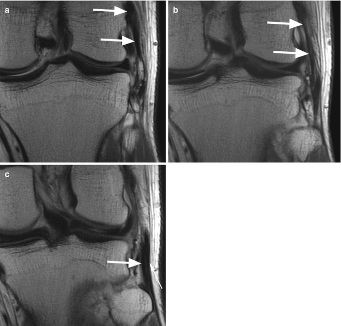

Fig. 4.1
Normal lateral collateral ligament (LCL) in a 21 year old male. Three consecutive coronal proton-density (PD) FSE images from anterior to posterior (a–c) show the normal LCL extending from the lateral femoral condyle to the fibular head (large arrows in a and b). At the fibular insertion LCL (large arrow in c) forms a conjoined tendon with the biceps tendon (small arrow in c)
The anterior oblique band (AOB) is a band of fibrous tissue that extends from the lateral collateral ligament to the lateral portion of tibia [11]. Some fibers of the anterior oblique band (AOB) blend with posterior fibers of the iliotibial tract [11].
On MR images, the lateral collateral ligament appears as a straight homogeneous hypointense structure on all sequences (Fig. 4.2). The anterior oblique band (AOB) is seen on axial MR images as a thin hypointense band extending from the lateral collateral ligament to the lateral tibia and the iliotibial tract (Fig. 4.3).
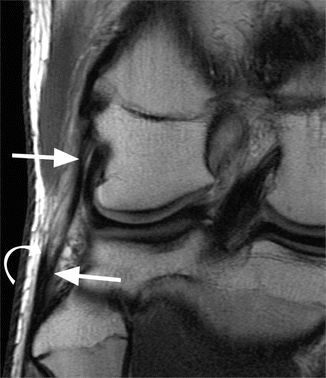
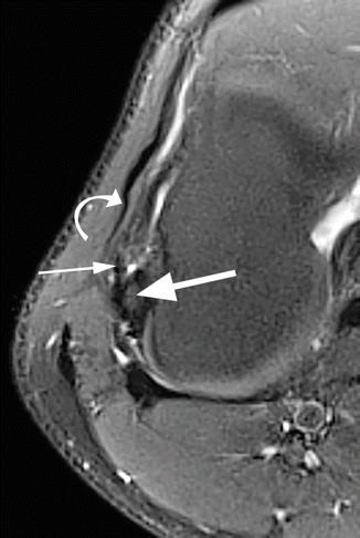

Fig. 4.2
Normal lateral collateral ligament (LCL) in a 22 year old male. In this case, the LCL is visualized on a single coronal proton-density (PD) FSE image as a continuous hypointense band (large arrows). At the distal insertion, the LCL and the biceps tendon (curved arrow) form a conjoined tendon

Fig. 4.3
Normal anterior oblique band (AOB) in a 17 year old male. Axial proton-density (PD) FSE fat-suppressed image shows the anterior oblique band (AOB) (small arrow) extending from the lateral collateral ligament (LCL) (large arrow) to the iliotibial tract (curved arrow)
4.1.2 Popliteal Tendon (PT), Anterolateral Ligament (ALL), Popliteomeniscal Fascicles (PMF), and Popliteofibular Ligament (PFL)
The popliteal tendon (PT) has its proximal attachment on the lateral femoral condyle, anteroinferiorly to the lateral collateral ligament. Two separate bundles are described at the proximal attachment: the posterior superficial bundle and the anterior deep bundle [12]. The popliteal tendon (PT) is intra-articular and extrasynovial at the level of the femorotibial joint and is surrounded by the popliteal bursa [13]. Distally, the tendon is extra-articular, deep to the fabellofibular ligament and the arcuate ligament [14]. It extends to the popliteal muscle. An extension of the synovial membrane between the posterior horn of the lateral meniscus and the popliteus tendon, known as popliteus or subpopliteus bursa, surrounds the tendon and may communicate with the superior tibiofibular joint (Fig. 4.4) [13]. On MR images, the tendon is hypointense on all sequences (Fig. 4.4). However, magic angle artifacts may occur due to its curved and oblique course especially at the proximal part. Thus, signal intensity irregularities should not be mistaken as tendinopathy or rupture per se but need to be verified on other sequences.
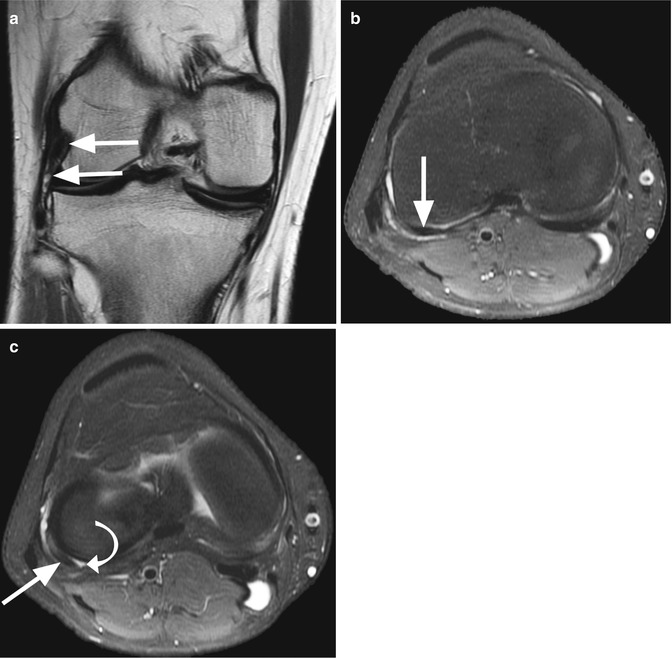

Fig. 4.4
Normal popliteal tendon (PT) and popliteus or subpopliteus bursa in a 19 year old female. Coronal proton-density (PD) FSE image (a) shows the hypointense PT at its femoral insertion (large arrows). On axial proton-density (PD) FSE fat-suppressed images (b, c), the PT is seen in contact with the lateral femoral condyle (large arrows in b, c). A small subpopliteus bursa (curved arrow in c) is seen between PT and posterior horn of lateral meniscus
The anterolateral ligament (ALL) is a relatively consistent structure that is found during knee arthroplasty [15]. It was first described as a reinforcement of the lateral capsule. The anterolateral ligament (ALL) takes origin from the lateral femoral condyle just anterior to and blending with the popliteus tendon, and it inserts distally to the lateral meniscus and lateral tibial plateau, typically about 5 mm distal to the joint line [15]. It can be seen on coronal MR images as a thin hypointense linear structure anterior to the popliteal tendon (PT) (Fig. 4.5).
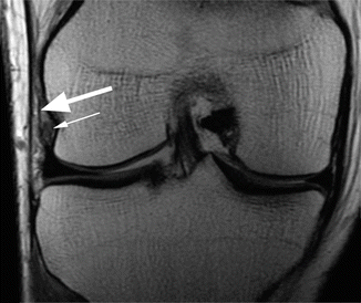

Fig. 4.5
The anterolateral ligament (ALL) in a 22 year old male. Coronal proton-density (PD) FSE image shows the ALL (large arrow) with its insertion on the lateral femoral condyle. The ligament blends with the popliteus tendon (small arrow)
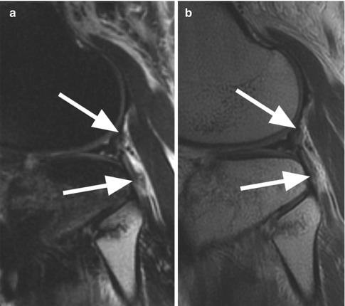
The popliteal tendon (PT) is strongly attached to the lateral meniscus through the two popliteomeniscal fascicles (PMF). The posterosuperior popliteomeniscal fascicle extends from the popliteal tendon to the posterolateral aspect of the lateral meniscus. The anteroinferior popliteomeniscal fascicle is stronger and shorter and extends from the popliteal tendon to the middle third of the lateral meniscus [12, 16]. On MR images, the popliteomeniscal fascicles are inconsistently seen as hypointense structures on sagittal planes (Fig. 4.6).
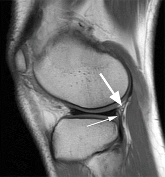

Fig. 4.6
The popliteomeniscal fascicles in a 19 year old female. Sagittal proton-density (PD) FSE image shows the posterosuperior popliteomeniscal fascicle (large arrow) and the anteroinferior popliteomeniscal fascicle (small arrow) strongly attached to the lateral meniscus
The popliteofibular ligament (PFL) attaches the popliteal tendon (PT) to the fibular head. It originates proximal to the myotendinous junction of the popliteus muscle and attaches to the medial fibular styloid [17]. The popliteofibular ligament (PFL) is approximately 10 mm long having a thickness similar to the lateral collateral ligament [12, 18]. The ligament is inconsistently identified on coronal MR images as a hypointense band (Fig. 4.7).
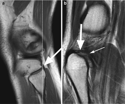

Fig. 4.7
The popliteofibular ligament (PFL) in a 19 year old female. Sagittal proton-density (PD) FSE image (a) shows the thick hypointense PFL (large arrow) inserting on the fibular head. On coronal proton-density (PD) FSE image (b) through the same level (line in a), the PFL (large arrow) is identified and connects the fibular head with the popliteal tendon (small arrow)
4.1.3 Arcuate Ligament (AL) and Fabellofibular Ligament (FFL)
The arcuate ligament (AL) is a Y-shaped thickening of the capsule with a medial and a lateral limb. The body of the arcuate ligament (AL) originates from the lateral edge of the styloid process of the fibula [19]. The lateral limb blends with the capsule and inserts to the lateral femoral condyle near the lateral gastrocnemius muscle (Fig. 4.8). The medial limb attaches to the posterior capsule.
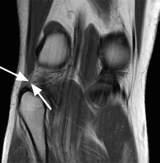

Fig. 4.8
The arcuate ligament (AL) in a 19 year old female. Coronal proton-density (PD) FSE image shows a thin hypointense band representing the lateral limb of AL (arrows) which originates from the lateral edge of the styloid process of the fibula and inserts to the lateral femoral condyle
The fabellofibular ligament (FFL) attaches proximally to the fabella and extends inferiorly and vertically to the fibular styloid process (Fig. 4.9). However, the fabellofibular ligament (FFL) may be present even in the absence of fabella [8, 20].
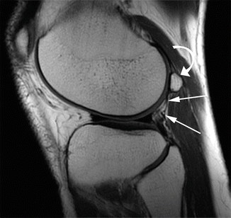

Fig. 4.9
The fabellofibular ligament (FFL) in a 35 year old male. Sagittal proton-density (PD) FSE image shows the FFL (arrows) which extends from the fabella (curved arrow) to the fibular styloid process on the fibular head. The fabellofibular ligament (FFL) may be present even in the absence of fabella
The arcuate ligament (AL) and the fabellofibular ligament (FFL) are not always present in anatomical studies. When present, the normal arcuate ligament (AL) and fabellofibular ligament (FFL) are depicted on MR images on coronal (arcuate ligament and fabellofibular ligament) and sagittal planes (fabellofibular ligament) as hypointense homogeneous bands (Figs. 4.8 and 4.9). However, in the authors’ experience, it remains difficult to identify both structures in the clinical routine imaging. One has to look specifically for these structures, and high-resolution images are mandatory for that.
4.2 MRI Pathological Findings
Although posterolateral corner injuries are not as common as injuries to the medial collateral ligament, they are more complex and more difficult to diagnose on physical examination. The most typical injury mechanism is a direct varus force to the anteromedial aspect of the hyperextended knee.
Almost all posterolateral corner lesions are associated with other injuries. The more common associated lesions are anterior and posterior cruciate ligament tears, medial collateral and medial meniscus injuries, anteromedial tibial plateau contusion or fracture, and the Segond fracture [1]. Untreated posterolateral injuries may be associated with chronic instability of the knee, failure of cruciate ligament reconstructions, and osteoarthritis [21, 22].
Appearance on MR images depends upon what structures are injured and the degree of injury. Lesions involving well-defined and well-delineated structures such as the lateral collateral ligament (LCL) and popliteal tendon (PT) can be classified on MR images into partial or complete tear. In the case of all the small structures mentioned above which, in addition, are inconsistently present and part of the capsule, the MR description should only refer to as “probably torn” based mainly on indirect signs.
4.2.1 Sprain and Partial Tears
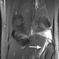
The sprain with intact fibers of the lateral collateral ligament (LCL) implies the presence of edema around the ligament with intact fibers (Fig. 4.10). A partial tear of the lateral collateral ligament (LCL) is seen on MR images as inhomogeneous signal intensity within the ligament. The ligament may be thinned or thickened without complete interruption of the fibers, and high-signal-intensity edema around the ligament is typically present (Figs. 4.11 and 4.12).
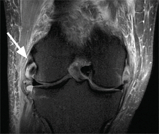
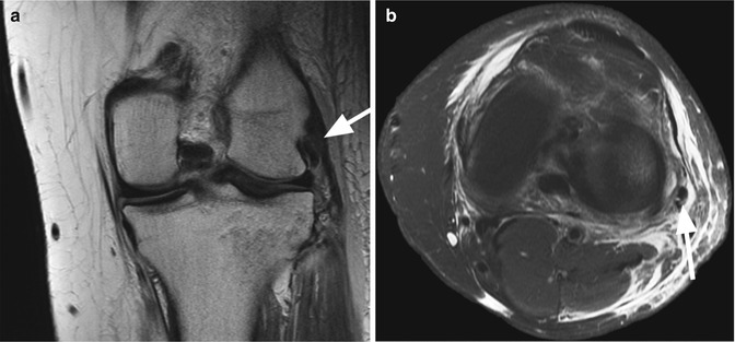
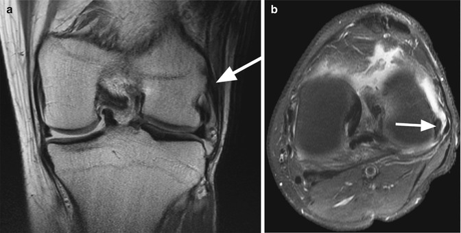

Fig. 4.10
Lateral collateral ligament (LCL) sprain in a 21 year old male. Coronal proton-density (PD) FSE fat-suppressed image shows extensive edema (arrow) around the proximal portion of LCL with intact fibers

Fig. 4.11
Lateral collateral ligament (LCL) partial tear in a 29 year old female. Coronal proton-density (PD) FSE fat-suppressed image (a) shows intrasubstance signal changes (arrow) at the proximal insertion of LCL without complete discontinuity of the ligament. The lesion is confirmed on axial proton-density (PD) FSE fat-suppressed image (b) where the tear is seen as a hyperintense linear signal lesion within the ligament (arrow)

Fig. 4.12
Lateral collateral ligament (LCL) partial tear in a 17 year old male. Coronal proton-density (PD) FSE fat-suppressed image (a) and axial proton-density (PD) FSE fat-suppressed image (b) show intrasubstance signal changes (arrow in a, b) at the proximal insertion of LCL without complete interruption of the fibers and high-signal-intensity edema around the ligament
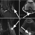
Most lesions of the popliteal tendon (PT) are extra-articular at the myotendinous junction. In the case of a partial tear of the popliteal tendon (PT), the injury is associated with edema and hemorrhage within the tendon and the musculotendinous junction. On MR images, the partial tear is seen as an amorphous or feathery signal intensity changes that may extend also into the muscle belly (Fig. 4.13).
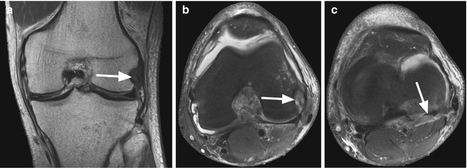

Fig. 4.13
Partial tear of the popliteal tendon (PT) in a 45 year old male. Coronal proton-density (PD) FSE fat-suppressed image (a) and axial proton-density (PD) FSE fat-suppressed image at the level of the femoral insertion (b) show intrasubstance signal changes (arrow in a, b). The lesion extension is usually better evaluated on serial axial images as in this case in which a more caudally axial (PD) FSE fat-suppressed image (c) shows the intrasubstance partial tear (arrow)
4.2.2 Complete Tears
In complete tears, there is a discontinuity of the involved anatomical structures that can be associated with waviness of the remaining ligament. In complete LCL tears, there is a discontinuity of the fibers, and edema or hemorrhage of high signal intensity on fluid-sensitive MR images is detected at the tendon defect (Figs. 4.14 and 4.15). A complete discontinuity of the popliteus musculotendinous junction with tendon retraction indicates a complete tear. When a hematoma is present, enlarged muscle volume and peri-fascial fluid collections are frequently seen [1]. In some cases, avulsion of the femoral insertion of popliteal tendon (PT) may be present.
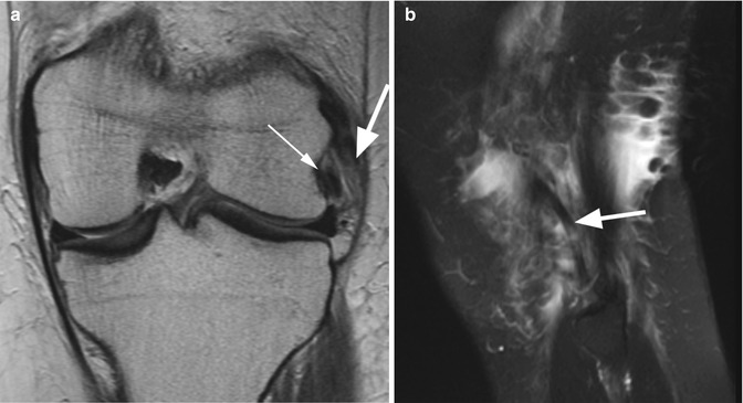
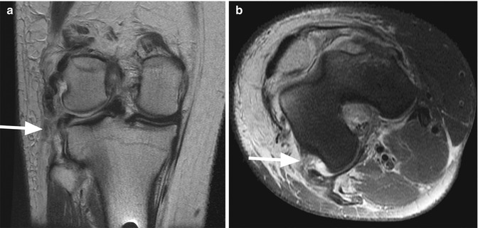

Fig. 4.14
Complete tear of the lateral collateral ligament (LCL) in a 25 year old soccer player. Coronal proton-density (PD) FSE image (a) shows discontinuity of the ligament (large arrow). Note the normal popliteal tendon (small arrow). Sagittal T2-weighted fat-suppressed image (b) demonstrates the interruption of the fibers (arrow) without a wavy contour of the ligament

Fig. 4.15
Complete tear of the lateral collateral ligament (LCL) in a 22 year old male. Coronal proton-density (PD) FSE fat-suppressed image (a) and axial proton-density (PD) FSE fat-suppressed image (b) show a complete tear of LCL with edema or hemorrhage at the site of the lesion (arrow in a, b)
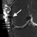
Individual assessment of the integrity of the anterolateral ligament (ALL), arcuate ligament (AL), popliteofibular ligament (PFL), and fabellofibular ligament (FFL) may not be possible [17]. MR signal abnormalities around the capsule, surrounding soft tissue edema or hemorrhage, and the lack of visualization of these structures are suggestive for popliteomeniscal fascicles (PMF) (Fig. 4.16), anterolateral ligament (ALL), arcuate ligament (AL), and fabellofibular ligament (FFL) tears. Normally, there should be fat tissue in this region which presents as homogeneous low signal intensity on fat-suppressed T2-weighted images (Fig. 4.17). Hyperintense signal on fat-suppressed T2-weighted MR images located posterior to the popliteal tendon (PT) suggests capsular tearing.

