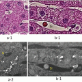
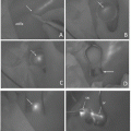
Fig. 26.1
(a) External view of HyperEye Medical System
(b) The conventional mode of IGFI (left side) and macroscopic view (right side) of intraoperative cholangiography
(c) The fusion IGFI mode. The IGFI image is fused to macroscopic image in a single monitor
26.2.2 Surgical Technique
Terminology surrounding the liver anatomy and resection is defined according to the International Hepato-Pancreato-Biliary Association, Brisbane, classification [14].
The indocyanine green (ICG) staining technique involved either systemic venous injection of a dye after clamping the inflow to the target vessels (IV method), portal puncture and direct injection (PV method), and systemic venous injection after clamping the outflow to the target major hepatic vein (vein-oriented staining method). Macroscopic observation under surgical lights was used to conduct the conventional demarcation technique.
Following a laparotomy with an inverted L-shaped incision, the liver was mobilized adequately according to the planned resection type. With the PV method, intraoperative ultrasonography (IOUS) was performed to confirm the tumor location and tumor-bearing portal branch.
26.2.3 IV Method
After dissection of the hepatic hilum, arterial and portal branches of the future resected hemiliver, section, or segment were exposed and taped. These inflow vessels were first temporarily clamped to confirm the demarcation line and intrahepatic blood flow by US, and then ligated and divided for a subsequent bolus intravenous injection of 2.5 mg ICG. Ten to twenty seconds after ICG injections, the splanchnic arteries and veins appeared enhanced on fusion IGFI, and ICG fluorescence was accumulated in the future remnant territory as counterstaining.
26.2.4 PV Method
After the puncture of the target portal branch, a mixture of 5 ml indigo carmine, 2.5 mg ICG, and 0.5 ml perflubutane suspension (Sonazoid; GE Healthcare, Oslo, Norway) was injected into the branch under contrast-enhanced (CE) IOUS guidance with the hepatic artery clamped. Under harmonic-imaging US guidance, we adjusted the injection speed according to the injection flow visualized in the portal vein to minimize the regurgitation or spillover into the non-eligible branches. The stained region was confirmed by macroscopic inspection, fusion IGFI, and CE-IOUS.
26.2.5 Vein-Oriented Staining
After full mobilization of the future resected liver, the target major hepatic vein was encircled and clamped at its root and 2.5 mg ICG was administered via bolus intravenous injection. After 3–4 min, the ICG had gradually accumulated in the non-veno-occlusive area, i.e., future remnant areas visible by counterstaining.
26.2.6 Acquisition of Fusion IGFI Images
After injection of a dye agent including ICG, video fluorescence images of the liver were recorded with the fusion IGFI camera positioned 40 cm above the subject surface and without surgical lights. Still fusion IGFI images could also be collected using the capture function of HEMS.
26.2.7 Acquisition of Conventional Demarcation Images
To gain conventional demarcation images, the near-infrared ray was turned off and macroscopic images were captured with the same function or with a digital camera under surgical lights.
26.3 Case Presentations
26.3.1 Case 1: IV Method
A 54-year-old woman developed intrahepatic cholangiocellular carcinoma located in the center of left lateral segment invading the umbilical portion, and we planned anatomical resection of left hemiliver.
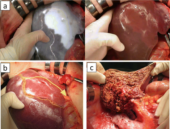
We performed IV method to gain the counterstaining of left hemiliver. After full mobilization of the left liver, hepatic hilum was dissected, and left hepatic artery and left portal vein were taped, respectively. IOUS was performed and inflow occlusion of left hemiliver was confirmed with selective clamp of both vessels. After ligation and division of these vessels, we administered 2.5 mg ICG intravenously. In 5 min, ICG was well accumulated in the remnant sections as counterstain, and clear demarcation appeared on the liver surface (Fig. 26.2a). Under Pringle maneuver, we performed parenchymal dissection using clamp crashing method. During parenchymal dissection, IGFI showed the intersegmental plane as the contour of parenchymal ICG uptake. During and after parenchymal dissection, we referred fusion IGFI monitor occasionally, judging whether ongoing dissection line is adequate or not (Fig. 26.2b, c, and d).
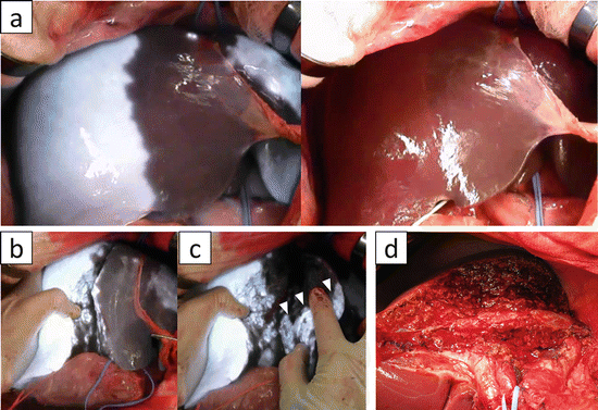

Fig. 26.2
Intrahepatic cholangiocellular carcinoma located in segment 3
(a) Fusion IGFI during counterstaining of left hemiliver using IV method (left side). The stained area is demarcated clearer than conventional demarcation method by vascular clamping (right side)
(b) Fusion IGFI during parenchymal dissection. The dissection plane is represented as contour of ICG uptake
(c) The deposit of ICG in specimen side (white arrowheads) indicates the dissection plane slightly extends toward the segment 5
(d) Macroscopic view after resection. The middle hepatic vein is longitudinally exposed as a landmark
26.3.2 Case 2: PV Method
A 52-year-old man developed hepatitis C-related HCC located in segment 8 and underwent anatomical resection of this segment.
After mobilization of the liver, intraoperative ultrasonography (IOUS) was performed, and each branch of portal pedicle in segment 8 was identified. We performed PV method targeting the main trunk of segment 8. In this case, the liver surface was poorly stained by conventional method, whereas fusion IGFI showed a clearer stained area than that of the macroscopic indigo carmine stain (Fig. 26.3a) and exactly corresponded to the demarcation line identified in the CE-IOUS and preoperative simulation.
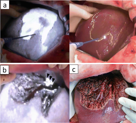

Fig. 26.3
Hepatitis C-related hepatocellular carcinoma located in segment 8
(a) Fusion IGFI of parenchymal staining of segment 8 using PV method (left side). The stained area is demarcated clearer than conventional indigo carmine staining (right side)
(b) After resection of segment 8. Fusion ICG indicates the small portion of dorsal area of segment 8 is remnant (arrowheads)
(c) Macroscopic view after resection. The right and middle hepatic vein is longitudinally exposed as a landmark of intersegmental plane
During parenchymal dissection, IGFI showed the intersegmental plane as the contour of parenchymal ICG uptake. After resection, we could evaluate the achievement of the anatomical resection by observing whether or not ICG fluorescence remained in the cut surface of the remnant liver (Fig. 26.3b, c).
26.3.3 Case 3: Vein-Oriented Staining Method
A 51-year-old man had developed synchronous multiple liver metastases from cecal cancer. Synchronous resection of primary lesion and liver metastases was considered. Preoperative and intraoperative imaging analysis showed 16 metastatic tumors located across the liver. Among them, 13 nodules were located within the watershed of the right hepatic vein (RHV). Volumetric analysis indicated that the veno-occlusive area of RHV corresponded to 40 % of total liver, and the liver functional reserve was well maintained. Thus, anatomical resection of the RHV watershed combined with limited resections in left liver was considered to be acceptable. After full mobilization of the right liver, the RHV was encircled and clamped at its root and 2.5 mg ICG was administered via bolus intravenous injection. After 3 min, the ICG had gradually accumulated in the non-veno-occlusive area, i.e., future remnant areas visible by counterstaining (Fig. 26.4a). We conducted parenchymal dissection according to the persistent and three-dimensional staining of the parenchymal border sacrificing the dorsal and lateral branches of segment 8 while preserving the ventral and medial branches, also extending to the segment 5 to clear the tumor margin (Fig. 26.4b). After resection, the watershed of RHV as well as segment 5 was extirpated completely, preserving the territory of ventral area in segment 8 (Fig. 26.4c).

