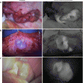
Fig. 42.1
Mechanisms of lymphangion for lymph transportation
42.2 Methods
42.2.1 Measuring the Human Lymph Pumping Pressure
All the studies described herein were approved by the Ethical Committee of Hamamatsu University School of Medicine, and informed consent was obtained from all the participants.
The measurement was performed as previously reported [9]. Briefly, using a 27-gauge needle, we subcutaneously injected 0.3 mL of ICG (Diagnogreen 0.5 %; Daiichi-Sankyo Pharmaceutical, Tokyo, Japan) at the dorsum of each participant’s foot. Immediately after the injection, fluorescence images of the subcutaneous lymphatic drainage were obtained using an infrared camera system (PDETM, Hamamatsu Photonics K.K. Hamamatsu, Japan), which activates ICG with light emitted at a wavelength of 760 nm and filters out light at a wavelength <820 nm. The light source for the emission of ICG consisted of 760-nm light-emitting diodes, and the detector was a charge-coupled device camera. The fluorescence images were continuously observed on the monitor of a laptop (VAIO, Type T; SONY Co., Tokyo, Japan).
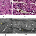
Before the injection of ICG, a custom-made transparent sphygmomanometer cuff was wrapped around the lower leg just below the popliteal fossa. The cuff was connected to a standard mercury sphygmomanometer. Lymphatic pumping was measured with the participants in the sitting position. Immediately after the subcutaneous injection of ICG, the transparent cuff was inflated to 70 mmHg, and then, it was gradually deflated to lower the pressure by increments of 5 mmHg every 2 min. This was performed until the fluorescence dye exceeded the upper border of the cuff, indicating that the lymphatic contraction had overcome the cuff pressure. The value of the cuff pressure at this point was used to measure P lymph pump (Fig. 42.2).
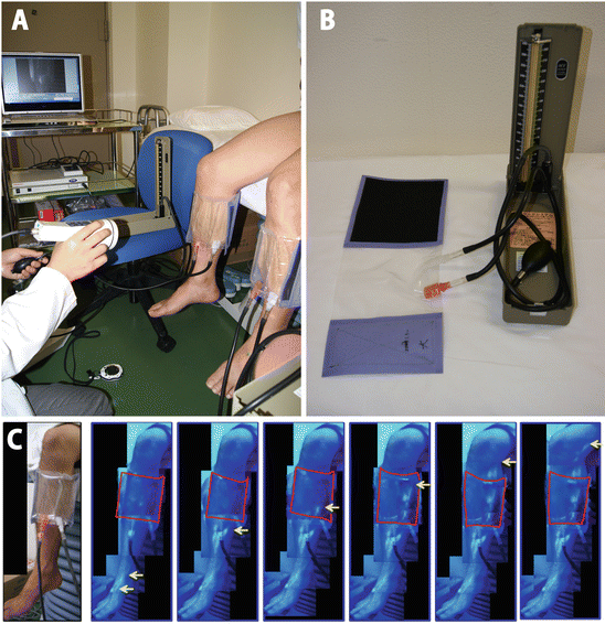
Fig. 42.2
(a) The measurement of lymphatic pumping pressure in the sitting position using PDE™ (Hamamatsu Photonics K.K. Hamamatsu, Japan)
(b) A custom-made transparent sphygmomanometer cuff and a standard mercury sphygmomanometer
(c) Real-time images of indocyanine green fluorescence lymphography after subcutaneous injection at the dorsum of the foot. The arrows (yellow) indicate the most advanced indocyanine green contrast agent in the lymph vessel; the solid line (red) outlines the transparent cuff (Cited from Ref. [9])
This method was confirmed by comparing data with the values obtained with a dynamic lymphoscintigram using the same cuff technique. We found that there was a significant correlation between the values with ICG fluorescence lymphography and dynamic lymphoscintigraphy [8].
42.2.2 The Effect of Aging on Human Lymph Pumping Pressure
Three hundred and ninety-nine participants (199 men and 200 women) were recruited from the Hamamatsu University School of Medicine. The subjects medical history was screened preliminarily, and those with no serious allergy and no history of leg injury or surgery were included. Those with an iodine allergy were excluded according to the industry’s recommendation because ICG contains iodine. In addition, subjects with varicose vein in the leg (i.e., C3, 4, and 5 according to the Clinical Etiology Anatomy Pathophysiology (CEAP) classification) were excluded. P lymph pump was measured as previously described.
Figure 42.3 shows that there was a significant correlation between the leg lymphatic pumping and age: r = −0.34 (p < 0.0001). Both sexes showed a significant correlation between leg lymphatic pumping and age (male: r = −0.32, p < 0.0001; female: r = −0.37, p < 0.0001).
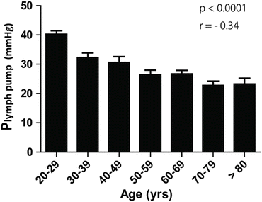
Fig. 42.3
The influence of age and sex on leg lymphatic pumping pressure (Plymph pump) (Cited from Ref. [12])
Aging affects the arteries and causes arteriosclerosis. However, venous stasis syndrome causes phlebosclerosis. Based on this study’s findings, aging may affect the lymph vessels and cause lymphosclerosis. This concept was proposed by Rabinovits et al. almost half century ago [10]. They performed a histological study on the human thoracic duct in elderly humans and reported changes in the distribution of smooth muscle cells and fibrosis. Our novel method of measuring enabled to demonstrate that “lymphosclerosis” may change the lymphatic vessels functionally. The mechanisms of contraction/relaxation in lymph vessels are yet to be fully understood. Similar to blood vessels, lymphatic endothelial cells have an ability to produce nitric oxide (NO) [11]. The endothelium-dependent modulation via NO production is considered to be a key mechanism of lymph pumping. In an animal study, endothelial NO synthase expression in the thoracic duct was markedly decreased, and flow-dependent modulation of the lymph pumping was completely abolished, suggesting that aging may disturb NO-dependent regulatory mechanisms of the lymphatic vessels [12].
Our study demonstrated the effect of aging on lymphatic vessels, which may begin a new era of lymphatic research regarding antiaging, lifestyle, and new drug developments [9].
42.2.3 Value of Human Lymphatic Pumping Pressure in Association with Leg Edema and Quality of Life
42.2.3.1 Methods
Four hundred and sixty-five healthy volunteers (78 men and 387 women; aged 30–85 years) participated in the study. A standard questionnaire was administered to obtain complaints of leg edema, body weight and height to calculate the body mass index (BMI), smoking status, and comorbid diseases (e.g., hypertension, diabetes mellitus, and hyperlipidemia). Complaints of leg edema were defined as a feeling of edema for at least 5 days per week even before noon. Subjects with varicose veins in the legs (C3, 4, and 5 according to the CEAP classification) or a medical history of lymphedema, deep vein thrombosis, radiation therapy, prescribed cancer agents, current pregnancy, or leg trauma were excluded.
The SF-36 was used to assess the participants subjective health conditions and their QOL. The SF-36 is a well-validated instrument that provides estimates for the following eight health concepts: physical functioning (PF); role functioning-physical (RP); bodily pain (BP); general health (GH); vitality (VT); social functioning (SF); role functioning-emotional (RE); and mental health (MH). Responses to the 36 questions represent these health concepts on a scale from 0 (worst possible health) to 100 (best possible health). Plymph pump was measured as described above.
42.2.3.2 Quality of Life with or Without Leg Edema
Approximately, 10 % of the participants complained of leg edema. There were no significant differences between those with and without edema with regard to BMI, smoking status, or comorbidities. The mean leg Plymph pump in the edema + and edema − groups were 20.4 ± 12.7 mmHg and 23.3 ± 15.6 mmHg, respectively (p = 0.23). There was no significant difference in the leg Plymph pump between the two groups, and there was no significant difference in age between the groups.
Related posts:
 ICG Fluorescent Image-Guided Surgery in Head and Neck Cancer
 Regional Lymph Node Dissection Assisted by Indocyanine Green Fluorescence Lymphography and Angiography for Stage III Melanoma
 Fluorescence Imaging for Intraoperative Identification of Pancreatic Leak
 Intraoperative Detection of Hepatocellular Carcinoma Using Indocyanine Green Fluorescence Imaging
 Basic Aspects of ICG Fluorescence Imaging of the Liver
 Principle and Development of ICG Method
Stay updated, free articles. Join our Telegram channel

Full access? Get Clinical Tree


