Magnetic Resonance Imaging Artifacts
Objectives
At the completion of this chapter, the student should be able to do the following:
Key Terms
An image artifact is a pattern or structure in the image caused by a signal distortion related to the technique of producing an image. Therefore it is an unwanted pattern or structure that does not represent the actual anatomy. Image artifacts can be misleading and interfere with diagnosis.
The complexity of magnetic resonance imaging (MRI) has unfortunately brought with it many confusing imaging artifacts. Luckily, many can be interpreted easily and do not interfere with diagnosis. However, the addition of each new imaging technique or pulse sequence brings the possibility of new artifacts.
The MRI artifacts illustrated here fall into several categories. The outline in Box 28-1 adopts a scheme of three principal classes of MRI artifacts, each of which can be identified as patient related or system related. None of these artifacts are characteristic of a particular manufacturer or model of MRI system.
Magnetic and Radiofrequency Field Distortion Artifacts
MRI requires an exceptionally uniform, homogeneous, primary magnetic field. Magnetic field homogeneity of ±1 ppm (parts per million) over a 20 cm spherical volume can be achieved by many current MRI systems.
Distortion of the magnetic field can be caused by the patient or problems with the MRI system. A poorly shimmed magnet, gradient magnetic field miscalibration, chemical shift of the Larmor frequency, tissue magnetic susceptibility, and foreign materials within or on the patient can all result in distortion of the magnetic field.
Patient Related
Foreign Materials.
Metals have different effects on the local magnetic field depending on their shape and location. Any metal is a potential conductor of electricity, which can support current induced by the pulsing of the gradient and RF magnetic fields. The currents produced by gradient coils will change the local magnetic field, leading to image distortions. The currents produced by the RF coils can lead to tissue heat and patient burns.
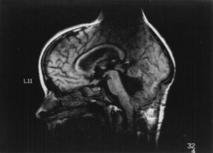
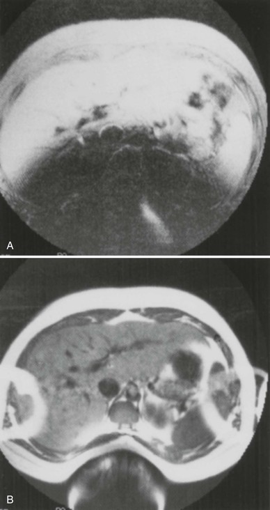
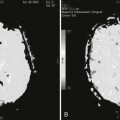
Ferromagnetic materials, in particular, should be avoided since they can establish magnetic fields that persist independent of the pulsing MRI secondary magnetic fields. Ferromagnetic materials can produce not only a local signal loss but also a warping distortion of the surrounding areas. An example of such field distortion is provided by the friendly “cone-head” seen in Figure 28-1.
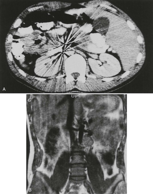
The typical metal material artifact has a partial or complete loss of signal at the site of the metal. Such metal can distort the local magnetic field sufficiently so that the Larmor frequency for local spins is outside the frequency range of the imaging system. Furthermore, metal contains no hydrogen; the result is signal void at that location.
Sometimes a partial rim of high signal intensity may be seen at the periphery of the signal void, which allows differentiation of metal from other causes of focal signal loss. Usually, the degree of anatomical information loss due to such an artifact is less than that seen on computed tomography (CT) images, because the loss is local and not streaked across the image as in CT.
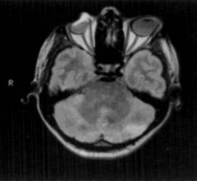
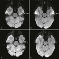
However, ferromagnetic material artifacts can actually cause very subtle, yet significant artifacts. The classic example is orthodontic braces, which cause severe signal dropout from the mouth and jaw. However, they can also cause artifacts in the remote regions in the brain by distorting the slice selection process.
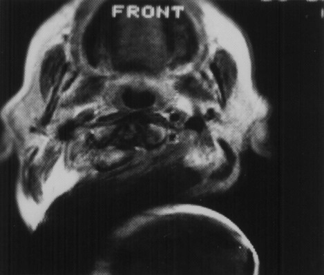
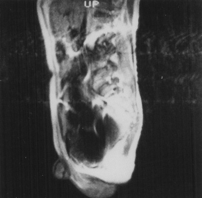
Exceptions to this are usually caused by the screws in some metallic orthopedic devices, but occasionally distortion is produced by small metallic objects such as buttons, snaps, zippers, or barrettes (Figure 28-2). The distortion of the surrounding tissue is the result of the metal-induced change in the local magnetic field. The magnetic field lines are distorted, resulting in a change in the local Larmor frequency (Figures 28-3 to 28-5).
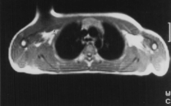
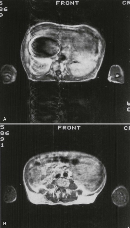
More mundane but relatively common metallic materials that may be encountered include belt buckles, keys, mascara and other makeup, foreign bodies in the eye or elsewhere, and even certain types of nylon found in clothing, such as gym shorts and warm-up suits (Figures 28-6 to 28-9).
Magnetic Susceptibility Artifacts.
Magnetic susceptibility is a dimensionless quantity that indicates the relative degree of magnetization occuring in a material that is placed in a magnetic field. Materials with magnetic properties that are different from the tissues in which they are embedded can cause a “susceptibility artifact.”
Stay updated, free articles. Join our Telegram channel

Full access? Get Clinical Tree


